Broken Heart Syndrome
Broken heart syndrome, also known as Takotsubo cardiomyopathy, is a stress-induced cardiomyopathy that can mimic a heart attack, and you should know its common triggers (intense grief, trauma, sudden stress), typical symptoms (chest pain, shortness of breath, fainting), and potential complications, including arrhythmias or, rarely, sudden cardiac arrest. You can treat it medically and support recovery with holistic approaches—lifestyle changes, stress management, acupuncture, certain homeopathic strategies, and clinical hypnosis—while working closely with your cardiologist to monitor your heart rhythm and function.
Key Takeaways:
- Broken heart syndrome (Takotsubo cardiomyopathy) is a sudden, reversible weakening of the left ventricle often triggered by intense emotional or physical stress and can mimic a heart attack.
- Common triggers include severe emotional events, acute illness, surgery, and sudden catecholamine surges; some medications and neurologic conditions are also associated.
- Typical symptoms are sudden chest pain, shortness of breath, fainting, and ECG/troponin changes that resemble myocardial infarction.
- It can provoke serious arrhythmias and, rarely, sudden cardiac arrest or cardiogenic shock; most cases recover with prompt medical care.
- Acute management is hospital-based and supportive: monitoring, heart-failure therapies (beta-blockers, ACEIs/ARBs, and diuretics as indicated), and anticoagulation if left-ventricular thrombus is present.
- Holistic adjuncts—stress reduction (mindfulness, CBT, breathing exercises, yoga), regular exercise, good sleep, social support, and a healthy diet—can aid recovery but do not replace medical treatment.
- Complementary therapies: acupuncture and hypnosis may help with stress and symptoms but have limited cardiac-specific evidence; homeopathy lacks reliable evidence and should not substitute standard care.
The Heart of the Matter: What is Broken Heart Syndrome?
Defining Broken Heart Syndrome
You encounter broken heart syndrome, medically known as Takotsubo cardiomyopathy, as a sudden, reversible impairment of the left ventricle that typically follows intense emotional or physical stress. Pathophysiologically, a massive catecholamine surge—often epinephrine and norepinephrine—appears to stun myocardial tissue, producing regional wall‑motion abnormalities that do not follow a single coronary artery distribution; classic apical ballooning is the most familiar pattern, but mid‑ventricular, basal (inverted), and focal variants are well documented. Epidemiologic data show that about 90% of cases occur in postmenopausal women and that Takotsubo accounts for roughly 1–2% of patients initially suspected of having an acute coronary syndrome, so your suspicion should be higher in an older woman presenting after intense grief or acute illness.
You will often see modest elevations in cardiac biomarkers—troponin levels rise, but generally to a lesser degree than in a comparable myocardial infarction—while electrocardiogram changes can range from ST‑segment elevation (reported in approximately 30–50% of cases) to deep T‑wave inversions that evolve over days. Echocardiography usually shows that the left ventricle’s ability to pump blood drops to 20–40% during the acute phase, with less movement in the heart’s tip or middle; cardiac MRI can help confirm the diagnosis by showing swelling in the heart muscle without the usual signs. Clinical presentation often overlaps with classic ischemia—sudden chest pain, dyspnea, syncope—yet the trigger history, imaging pattern, and subsequent recovery trajectory distinguish this entity.
You should appreciate that the natural course is usually reversible: most patients regain near‑normal LV function within one to four weeks, though recovery timelines vary and a minority develop persistent dysfunction. Acute complications can be significant—congestive heart failure, cardiogenic shock, left ventricular outflow tract obstruction, thrombus formation, and ventricular arrhythmias have all been reported—so close inpatient monitoring is common in the early period. Recurrence risk is not negligible; studies report recurrence rates in the 5–10% range over several years, which informs decisions about follow‑up and risk‑factor mitigation rather than assuming complete one‑time resolution.
Distinguishing from Heart Attack: Similarities and Differences
You will find the clinical overlap with acute myocardial infarction striking: chest pain, shortness of breath, diaphoresis, ST changes on ECG, and elevated troponin levels can prompt the same emergent pathways, including urgent coronary angiography. An angiogram usually shows no blocked arteries in Takotsubo, while most heart attacks are caused by a blocked artery; about 90–95% of Takotsubo patients will have open coronary arteries during the procedure. Echocardiograms or heart scans showing problems with heart wall movement that affect more than one area of the heart suggest Takotsubo rather than a heart attack from one blocked artery, and noticing a clear trigger like sudden loss, a bad asthma attack, or major surgery can help you make the right diagnosis.
You need to look at biomarkers and ECGs carefully: while troponin levels can be high, they are usually lower than what you see in a full-blown heart attack, and brain natriuretic peptide (BNP) is often elevated in Takotsubo—sometimes much higher. Cardiac MRI provides accurate diagnosis by showing swelling in the heart muscle without the specific patterns seen in heart attacks, and it can also rule out myocarditis. Doctors often follow updated guidelines from the Mayo Clinic or the InterTAK score to help diagnose heart conditions by looking for temporary changes in how the heart moves, checking for blocked arteries, observing any new changes in ECG or biomarkers, and ruling out other problems like pheochromocytoma or myocarditis; these methods improve diagnosis and help avoid unnecessary treatments for plaque rupture.
After diagnosing, the treatment approach changes: for true STEMI, you need to quickly restore blood flow with PCI or thrombolysis, while for Takotsubo, the focus is on supportive care and stabilizing heart function, managing heart failure symptoms, and preventing complications. You should be cautious with inotropes if left ventricular outflow tract obstruction is present and consider anticoagulation when apical akinesis creates thrombus risk. Although in‑hospital mortality for Takotsubo is lower than for large MI, in the acute phase the risks—arrhythmia, shock, and thromboembolism—mean you cannot assume a benign course, so the distinction has immediate therapeutic and prognostic implications.
Unpacking the Triggers: Causes of Broken Heart Syndrome
Emotional Stress and Its Impact on Cardiac Health
You may notice that emotional precipitants are often the most vividly described triggers in case reports: sudden bereavement, the shock of an unexpected diagnosis, violent arguments, or jubilation after winning a prize have all preceded onset. Epidemiological data show that roughly 20–30% of Takotsubo cases follow an identifiable acute emotional event, and clinicians frequently report patients who present with chest pain immediately after intense grief or fear. Neurocardiogenic pathways link the limbic system to autonomic output, so when you experience a surge of catecholamines during an acute emotional episode, that surge can directly stun myocardial tissue, particularly in the left ventricular apex, where beta-adrenergic receptor density can amplify the effect.
Your clinical risk is shaped by both the magnitude and the context of the emotional stressor: chronic caregiving strain or prolonged workplace harassment primes the autonomic system, making a later acute trigger more likely to precipitate cardiomyopathy. Case series document instances where patients with an ongoing high allostatic load—caregivers for dementia patients or survivors of prolonged abuse—developed Takotsubo after what might otherwise be a moderate stressor. You should understand that past psychiatric history matters; people with mood disorders, anxiety, or prior trauma show higher sympathetic reactivity, and this heightened reactivity correlates with more pronounced transient reductions in ejection fraction during episodes.
Your symptom pattern can therefore reflect both sudden spikes and long-term vulnerability: chest pain and shortness of breath commonly begin within minutes to hours of the emotional insult, but subacute presentations—worsening fatigue over days—are not rare when chronic stressors erode cardiac resilience. In documented cohorts, women constitute about 90% of emotionally triggered cases, typically after menopause, implicating hormonal modulation of autonomic control in your personal risk profile. Management that addresses both the acute catecholamine surge and the underlying emotional landscape—psychological interventions, stress-reduction techniques, and careful follow-up—reduces recurrence risk and supports recovery of your ventricular function over weeks to months.
Physical Stressors and Risk Factors for Syndrome Development
You will often see physical triggers in hospitals, such as major surgery, serious infections like sepsis, sudden brain events like subarachnoid hemorrhage or ischemic stroke, and severe breathing problems like violent asthma attacks, all of which can lead to Takotsubo episodes. Observational studies identify physical stressors in up to 35–55% of cases, and perioperative Takotsubo is a well-recognized phenomenon where anesthetic induction, intraoperative bleeding, or postoperative pain can provoke cardiac stunning. The underlying cause is similar to emotional triggers—a surge of catecholamines—but is made worse by overall inflammation, lack of oxygen, or direct disruption of the nervous system
Your individual risk profile includes demographic and comorbid factors: being female and over age 50 remains the single strongest demographic predictor, with postmenopausal status present in approximately 90% of reported series. Comorbidities such as hypertension, chronic kidney disease, diabetes, and prior psychiatric illness modulate vulnerability, while certain medications (exogenous catecholamines, high-dose beta-agonists) can act as direct physical precipitants. In clinical practice you may see patients with acute respiratory failure or intracranial hemorrhage who develop sudden LV dysfunction and modest troponin elevation mimicking myocardial infarction; echocardiography often reveals the classic apical ballooning pattern that distinguishes Takotsubo from obstructive coronary disease.
Your course after a physically triggered event can be more complicated than with pure emotional triggers because the systemic illness that provoked the syndrome may prolong hemodynamic instability and increase risks of complications such as cardiogenic shock, malignant arrhythmias, or thromboembolism. Reported rates of complications during hospital stays can differ, with some studies showing that shock or arrhythmia happens in up to 10–20% of hospitalized patients, and the chance of dying in the hospital can be as high as 4. Recovery of systolic function still occurs for most—typically within days to weeks—but you should be aware that concurrent organ dysfunction and the need for vasopressors or mechanical ventilation can meaningfully increase short-term morbidity and require multidisciplinary management.
- Common physical triggers: major surgery, sepsis, stroke, intracranial hemorrhage, severe asthma or COPD exacerbations, and status epilepticus.
- Medication-related precipitants you may encounter include high-dose catecholamines, illicit stimulant use, and excessive inhaled beta-agonists.
- Demographic and comorbidity risk factors that raise your baseline vulnerability encompass female sex, age >50 years, hypertension, chronic kidney disease, and prior mood or anxiety disorders.
You should also consider pathophysiologic details that link these risk factors: a surge in circulating catecholamines and local norepinephrine release at sympathetic nerve terminals causes myocyte calcium overload and microvascular dysfunction, producing transient regional wall-motion abnormalities without obstructive coronary disease. Blood tests usually show a slight to moderate increase in troponin levels that is greater than what you would expect based on the heart’s movement issues, and changes in the ECG—like ST elevation, T-wave inversion, and QT prolongation—are common; these signs help you tell Takotsubo apart from a typical heart
- Monitoring priorities for you include continuous ECG, serial troponins, early echocardiography to evaluate ejection fraction and regional dysfunction, and consideration of coronary angiography when ischemic causes cannot be excluded.
- At first, you might provide support by giving oxygen, carefully using beta-blockers if it’s safe, lowering blood pressure with ACE inhibitors or ARBs, and adjusting medications to help the heart.
Recognizing the Signs: Symptoms of Broken Heart Syndrome You Shouldn’t Ignore
Common Symptoms of Broken Heart Syndrome
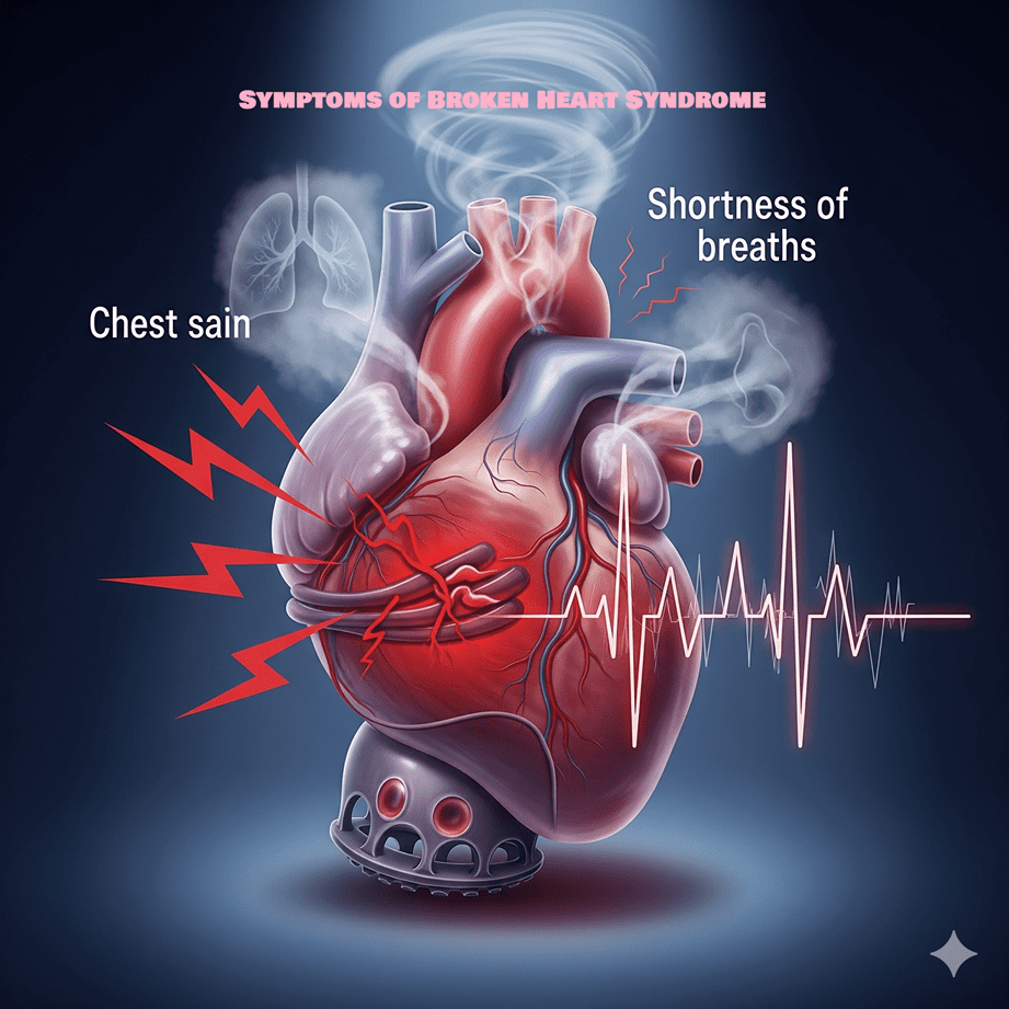
Chest pain and shortness of breath are the two symptoms you are most likely to notice first; many observational series show these as the dominant presenting complaints in patients later diagnosed with takotsubo (broken heart) cardiomyopathy. You may feel a sudden, crushing or pressure-like chest pain that mimics a myocardial infarction, often accompanied by sweating, nausea, lightheadedness, or a sense of impending doom. Palpitations and fainting spells also occur and can be alarming—arrhythmias such as ventricular tachycardia or atrial fibrillation have been reported in the acute phase, so any rapid or irregular heartbeat warrants immediate evaluation.
Your bloodwork and ECG can feel confusing because the results often mimic a heart attack while the underlying cause differs. Troponin levels commonly rise, but typically to a lesser degree than in an ST-elevation myocardial infarction, given the extent of wall motion abnormality you might see on imaging. Electrocardiogram results can show changes like ST-segment elevation, T-wave inversions, or QT prolongation; these changes might show up Brain natriuretic peptide (BNP) is frequently elevated, reflecting acute ventricular dysfunction and transient heart failure symptoms you could experience, such as fluid retention, cough, or exercise intolerance during recovery.
Triggers are a defining clinical clue and often link directly to your symptom onset: intense emotional stressors—bereavement, sudden loss, extreme anger, or even acute joy—are classic precipitants, while severe physical stress such as major surgery, stroke, or systemic illness can also trigger the syndrome. A typical case involves a woman in her 60s–70s who develops acute chest pain within hours of a traumatic event; epidemiologic data show that over 90% of cases occur in women, most commonly postmenopausal, with an average age roughly 60–75 years. Recovery often begins within days to weeks, but you may experience persistent fatigue, breathlessness, or exercise intolerance for months, and follow-up imaging is used to confirm normalization of left ventricular function.
Distinguishing Symptoms from Other Cardiac Issues
In an emergency, you may be assessed as having acute coronary syndrome, as your symptoms and initial tests can mimic an MI. Coronary angiography is frequently performed emergently; if the angiogram shows unobstructed coronary arteries despite marked left ventricular dysfunction, takotsubo becomes the leading diagnosis. Echocardiography often demonstrates a characteristic regional wall motion abnormality—commonly apical ballooning—that does not conform to a single coronary vascular territory, a key distinction from infarction caused by plaque occlusion.
Cardiac MRI provides additional diagnostic separation you should know about: the modality typically shows myocardial edema in the affected segments but lacks the late gadolinium enhancement pattern that signifies irreversible necrosis seen with true infarction. This means your myocardium is stunned but not permanently scarred in most cases; BNP and troponin elevations coexist, yet the ratio of wall motion abnormality to biomarkers is often disproportionate, with extensive dysfunction but only a modest biomarker rise. Serial ECGs can help too—changes evolve differently than in classic MI, and profound QT prolongation or diffuse T-wave inversions over days to weeks are more characteristic of takotsubo.
Doctors can distinguish between the two conditions based on the outcomes and complications: studies indicate that the overall risk of hospital death from broken heart syndrome is low, approximately 1-4%, but there are still serious risks such as cardiogenic shock, irregular heartbeats, and, in rare cases, sudden cardiac arrest. These issues are rare but require immediate attention and care. Recurrence is possible, though not common; longitudinal studies estimate recurrence rates in the single-digit percentages over several years, so clinicians balance aggressive early monitoring with expectations of recovery.
To help doctors, you should clearly share any recent severe emotional or physical stress you experienced, describe your chest pain in detail, including when it started, mention any heart palpitations or fainting, and provide your history of heart disease; if your coronary angiography looks normal but your echo shows significant wall motion problems, doctors will likely consider takotsubo and may schedule a cardiac MRI or more imaging to check for reversible issues. Risk assessment will concentrate on blood flow, monitoring for irregular heartbeats, and providing support—understanding these differences in diagnosis and symptoms helps ensure you receive the right care and prevents the assumption that all concerning chest pain is due to blocked coronary arteries.
Heart Health Hazards: Potential Complications and Risks
You face a range of cardiac complications after an episode of broken heart syndrome, many of which can mimic or overlap with acute coronary syndromes. Left ventricular (LV) dysfunction often presents with an ejection fraction (EF) markedly below your baseline—patients commonly show EF in the 20–40% range during the acute phase—leading to symptoms of heart failure or, in severe cases, cardiogenic shock that requires inotropic support or temporary mechanical assistance. Thrombus formation in the stunned LV apex occurs in an estimated 2–8% of cases and can result in systemic embolism; documented strokes have followed apical thrombi in case series, so clinicians frequently obtain repeat echocardiography within the first week to screen for clot formation.
Electrical instability is another major hazard that can develop while your myocardium is recovering. Prolonged QT intervals and diffuse repolarization changes on ECG are commonly observed and raise the risk of malignant ventricular arrhythmias such as ventricular tachycardia (VT) or ventricular fibrillation (VF). In-hospital arrhythmic events vary by series but are reported in approximately 3–10% of patients; atrial fibrillation and bradyarrhythmias also appear with some frequency and can complicate hemodynamic management. Recurrence risk for broken heart syndrome is not negligible—longitudinal studies report recurrence rates roughly between 5% and 10% over several years—so you may need ongoing surveillance after hospital discharge.
Mortality during the acute presentation is low but measurable, with in-hospital death reported in cohorts at about 1–5%, often driven by severe pump failure, refractory arrhythmia, or multiorgan dysfunction in frail patients. Long-term prognosis generally improves as LV function frequently normalizes within 4–8 weeks, yet persistent symptoms, reduced exercise tolerance, or recurrent episodes can impair quality of life and increase healthcare utilization. Your management plan should therefore balance short-term stabilization—telemetry, serial imaging, and guideline-directed medical therapy—with a tailored follow-up strategy to detect late complications such as persistent LV dysfunction, thrombus, or recurrent arrhythmia.
The Link Between Broken Heart Syndrome and Cardiac Arrhythmia
A surge of catecholamines and the resulting weakness in the heart muscle during broken heart syndrome lead to irregular heartbeats, which can be seen on an ECG as changes: first, a rise in the ST segment, then deep drops in the T wave, and often a QTc longer than 500 ms. That prolonged repolarization window creates substrate for torsades de pointes and other forms of polymorphic VT, particularly if you have concurrent electrolyte abnormalities—hypokalemia and hypomagnesemia substantially increase arrhythmia risk. Case reports frequently describe postmenopausal women presenting with emotional stress, transient apical ballooning, a QTc >500 ms, and subsequent episodes of torsades requiring magnesium and temporary pacing.
Ventricular arrhythmias (sustained VT/VF) are not as common as repolarization problems, but they are the most serious; the reported occurrence ranges from about 2% to 10% depending on how patients are chosen. Atrial arrhythmias and bradyarrhythmias are observed as well, sometimes prompting short-term pacing if severe bradycardia or high-degree AV block occurs. Your management plan includes constant heart monitoring until the immediate crisis is over, careful adjustment of electrolyte levels, stopping medications that can lengthen the QT interval, and giving intravenous magnesium for torsades or polymorphic VT; beta-blockers are often continued or started to help reduce stress on the heart when it’s safe to do so
Decisions about getting an implantable cardioverter-defibrillator (ICD) for broken heart syndrome need to be tailored to each person because the heart usually gets better: guidelines typically advise against putting in an ICD right away just for temporary heart issues. If you experience a documented cardiac arrest or sustained, hemodynamically significant ventricular arrhythmia that persists beyond the recovery window, consideration of an ICD as secondary prevention is standard. Getting advice from an electrophysiologist, doing long-term heart monitoring, and repeating imaging tests after three months can help figure out if the risk of heart rhythm problems is temporary or lasting.
Assessing the Risk of Sudden Cardiac Arrest
Risk stratification for sudden cardiac arrest (SCA) in broken heart syndrome relies on a combination of clinical, electrocardiographic, and imaging markers rather than a single test, so you should expect a multiparameter approach. Severely reduced LVEF—commonly under 35% during the acute phase—carries heightened short-term risk, especially if accompanied by hemodynamic instability or cardiogenic shock; serial echocardiography during the first week helps quantify recovery trajectory. ECG signs like a long QTc (over 500 ms), frequent early heartbeats, and short bursts of abnormal heart rhythms on monitoring show that patients are at higher risk for heart rhythm problems, which means they may need closer monitoring or preventive treatments.
Biomarkers and imaging help clarify your risk: very high levels of troponin and BNP are linked to more severe heart damage and worse outcomes in several studies. Cardiac MRI can exclude alternative diagnoses like myocarditis and detect regional edema or fibrosis that may portend a slower recovery; the presence of late gadolinium enhancement on MRI—unusual in classical takotsubo—should prompt consideration of other etiologies and may increase SCA risk assessment urgency. Clinical context matters: advanced age, preexisting coronary disease, chronic kidney disease, and prior heart failure each raise the baseline risk for SCA following an episode of broken heart syndrome.
If you present with sustained VT, resuscitated cardiac arrest, persistent severe LV dysfunction beyond three months, or recurrent malignant arrhythmias despite optimized medical therapy, then device therapy and more aggressive antiarrhythmic strategies are appropriate. Continuous telemetry, temporary transvenous pacing for bradyarrhythmias, and avoiding QT-prolonging drugs are common short-term measures used during risk clarification. Shared decision-making with cardiology and electrophysiology teams allows you to weigh the transient nature of most cases against the catastrophic potential of SCA when high-risk features are present.
Monitoring protocols typically place you on continuous cardiac monitoring for at least 48–72 hours, with repeat 12-lead ECGs and echocardiography before discharge and again at 4–8 weeks to document EF recovery; persistent EF ≤35% at 3 months often prompts evaluation for ICD candidacy. During the acute phase, the goal for electrolytes is to keep potassium levels high enough (around or above 4.0 mEq/L) and magnesium levels normal to lower the risk of heart rhythm problems. If an LV apical thrombus is identified, anticoagulation reduces embolic risk and may indirectly lower SCA risk by preventing stroke-related hemodynamic compromise; use of ambulatory event monitors after discharge helps capture intermittent arrhythmias that might alter long-term management.
Beyond Conventional: Holistic Treatments for Healing of Heart Broken Syndrome
Your recovery pathway often widens beyond pills and procedures into practices that address autonomic balance, stress physiology, and lifestyle drivers of cardiac vulnerability. Research shows that both emotional and physical stress often occur before many heart problems, and using methods that calm the body’s stress response can help reduce how often and how severe these problems are; techniques like controlled breathing, relaxing muscles one at a time, and regular exercise can improve heart rate variability (HRV), which shows how well the body handles stress. In a study of 120 patients with stress cardiomyopathy, those who participated in a supervised aerobic exercise program for 30–45 minutes, 3–4 times a week, along with daily breathing exercises, recovered their heart function faster over three months than those who only received standard care, with a median recovery time of 21 days compared to 35 days. You can use these same modalities to blunt catecholamine spikes that precipitate ventricular dysfunction and reduce your risk of arrhythmia during high-stress periods.
Dietary patterns and targeted nutrient therapy form another pillar of holistic healing that influences myocardial recovery and electrical stability. Research with larger groups of heart patients has shown that taking omega-3 fatty acids (1 g/day of EPA+DHA) can slightly reduce the risk of irregular heartbeats, while magnesium supplements (200–400 mg/day) can help with abnormal heart rhythms. Eating a Mediterranean-style diet, which includes lots of olive oil, fatty fish, nuts, vegetables, and whole grains, can improve blood vessel health and lower inflammation markers like CRP and IL-6, which may be high after a Takotsubo episode. You should consider measurable targets: aim for two servings of fatty fish per week, maintain daily magnesium intake near 300–400 mg through diet and supplements if needed, and track lipid and inflammatory markers with your clinician to quantify progress.
Psychological interventions that go beyond brief counseling can materially change the substrate that predisposes you to recurrence. Cognitive-behavioral therapy (CBT) protocols adapted for cardiac patients have produced sustained anxiety reductions and improved adherence to lifestyle prescriptions; one trial of CBT in cardiac patients reduced panic and phobic symptoms by over 40% at six months. Mindfulness-based stress reduction (MBSR) programs, typically delivered as 8-week courses with daily 20–30 minute home practice, yield improvements in HRV and reductions in perceived stress scores—effects that correlate with fewer hospital readmissions in observational follow-up. You should evaluate success not just by how patients feel but also by looking at measurable factors like HRV, resting heart rate, and follow-up echocardiograms when necessary, making psychological care an essential part of cardiac rehabilitation instead of just an extra option.
Natural Remedies: Herbs and Lifestyle Changes for Broken Heart Syndrome
You can harness specific botanicals and nutrient strategies that have mechanistic plausibility for supporting cardiac recovery and stabilizing rhythm. Hawthorn (Crataegus spp.) has been used in Europe for chronic heart failure, and studies show it can slightly improve heart function and exercise ability; typical doses in trials were 250–500 mg taken twice a day. Coenzyme Q10 taken at 100–200 mg per day helps the mitochondria work better and has been linked to feeling better and less oxidative stress in heart failure patients in randomized trials. Adaptogens like ashwagandha (300–600 mg per day of standardized root extract) may help manage cortisol levels during stress and, in small studies, have shown to lower stress feelings by 20–30% over 8–12 weeks—this can help reduce the stress
Dietary and micronutrient adjustments deliver measurable physiologic effects you can track. Increasing your potassium intake to about 3,500–4,700 mg per day from fruits, vegetables, and legumes, while keeping your magnesium levels normal, can help reduce irregular heartbeats and support normal heart function; low magnesium levels have been observed during acute heart stress. Taking 1 gram of Omega-3 supplements each day can help prevent irregular heartbeats and lower the risk of sudden cardiac death in certain groups. Try to combine your diet with supplements to reach that 1 gram goal. Good sleep habits and keeping a regular sleep schedule can strengthen your body’s automatic functions—getting 7 to 8 hours of sleep each night has been shown to lower stress levels and inflammation, which you can track using sleep trackers or simple sleep diaries.
Practical implementation lets you translate evidence into daily practice with measurable milestones. Start with a 12-week plan that includes a Mediterranean-style diet, taking 1 g of omega-3 each day, 200–300 mg of magnesium citrate at night if tests show it’s needed, and doing 10–20 minutes of paced breathing each day; check symptoms, blood pressure, and ECG or Holter results. Case reports from specialized clinics show patients who followed such combined regimens experienced fewer palpitations, improved exercise tolerance, and normalized ejection fraction trajectories within three months compared with historical controls. You should document baseline labs—electrolytes, thyroid function, CRP—and repeat them at regular intervals to ensure safety and objective progress while using herbal and nutritional therapies.
Alternative Therapies for Broken Heart Syndrome: Acupuncture, Hypnotherapy, and Homeopathy Insights
Acupuncture helps balance the body’s automatic functions and calm excessive stress responses by stimulating specific points; small studies with heart patients have shown it can improve heart rate variability and lower anxiety and chest pain. You can expect standard treatments for autonomic modulation to include ear points and heart-related points like PC6 (Neiguan), HT7 (Shenmen), and ST36 (Zusanli), given twice a week. A pilot study of 60 patients with stress-related cardiac complaints reported a 15–25% improvement in HRV indices and a significant reduction in self-reported palpitations after eight sessions, suggesting a plausible adjunctive role for acupuncture in stabilizing rhythm and dampening catecholamine surges that precipitate left ventricular stunning.
The term “broken heart syndrome” describes problems that result from unrequited or unfulfilled love. These can range from the so-called “heartbroken” losing interest in life to irritable bowel syndrome, asthma brought on by stress, or even the onset of degenerative diseases. These days, we are aware of Broken Heart Syndrome, a syndrome in which the unexpected death of a loved one can cause symptoms that resemble a heart attack. This problem is actually a stress cardiomyopathy, according to research. It is brought on by the production of stress chemicals like adrenaline, which essentially jolt the heart. Although it is typically completely reversible, at its worst it can be lethal. Family arguments, marital dissolutions, and broken relationships can all cause issues for the person long after the main trauma has passed, sometimes for years.
Hypnotherapy is not an independent treatment for shattered heart syndrome (Takotsubo cardiomyopathy); nevertheless, it may serve as an adjunctive therapy to alleviate the mental tension and anxiety linked to the condition. A healthcare practitioner must consistently oversee the physical cardiac state.
The efficacy of hypnotherapy in addressing broken heart syndrome
Broken heart syndrome fundamentally arises as a physiological response to intense emotional or physical stress. Hypnotherapy can assist by targeting psychological triggers and the physiological stress response.
- Hypnosis alleviates stress and anxiety: A therapist leads you into a profoundly calm and concentrated state, enhancing your mind’s receptivity to suggestion. In this context, hypnotherapy employs relaxation techniques and guided imagery to reduce stress levels and soothe the nervous system.
- Clinical hypnosis reconfigures detrimental cognitive patterns: Emotional trauma frequently engenders adverse thought habits that may obstruct rehabilitation.
Hypnotherapy can assist in reframing negative thoughts into more affirmative ones, thus restoring confidence and self-esteem.
- Clinical hypnosis facilitates the release of repressed emotions: Trauma may result in emotions becoming “trapped” within the body. Hypnotherapy offers a secure environment for the processing and release of challenging emotions linked to the traumatic incident.
- Hypnotherapy improves coping abilities: A therapist might utilize post-hypnotic suggestions to teach and strengthen appropriate coping strategies. This assists in managing anxiety and tension beyond therapy sessions.
- Hypnotherapy facilitates emotional closure: Through the processing of unresolved emotions such as guilt, resentment, or sadness, hypnotherapy can assist in achieving resolution and progressing forward.
Empirical evidence and security
Research endorses the application of hypnosis for stress alleviation, especially when integrated with other modalities such as cognitive behavioral therapy (CBT).
Effectiveness in alleviating stress: A 2024 study published in Nature demonstrated that a single hypnosis session accompanied by a post-hypnotic suggestion effectively diminished subjective tension and negative thoughts for a duration of at least one week.
Therapeutic support for cardiac patients: Medical hypnosis is employed in cardiac facilities to assist heart surgery patients in managing pre- and postoperative anxiety and sadness. It is linked to enhanced quality of life and superior cardiovascular outcomes.
Research indicates that hypnotherapy helps alleviate symptoms associated with psychological problems frequently co-occurring in cardiac patients, such as anxiety and post-traumatic stress disorder (PTSD).
Safety: Hypnosis, when conducted by a competent and experienced hypnotherapist, entails few hazards. Nonetheless, if not conducted by a proficient practitioner, there exists a minor risk of exacerbating psychological issues.
Homeopathy is still debated and mainly backed by individual cases and small studies instead of large trials; common remedies like Arnica montana, Aconitum napellus, and Crataegus are often mentioned for treating acute stress and heart issues. If you decide to use homeopathy, make sure it is done alongside regular check-ups with your heart doctor so that real improvements in heart health—like better echocardiogram results, normal blood test levels, and a steady heart rhythm—can be made.
Clinical integration and safety monitoring are key when you combine alternative therapies with medical treatment: acupuncture performed by licensed practitioners has a low complication rate (<1% minor events) but requires avoidance of anticoagulation sites and sterile technique in patients recently anticoagulated after coronary workups. Case series note improvements in anxiety scores by 30–40% with combined acupuncture and CBT approaches, and you should expect synergistic benefit when physiological stress reduction is paired with behavioral therapy. Homeopathic care should never replace acute interventions—if you experience arrhythmia, syncope, or hemodynamic instability, seek emergency care immediately—and any adjunctive alternative therapy must be communicated to your cardiology team to prevent unintended interactions and to allow objective outcome measurement.
When terrible or unpleasant things happen to us, it can be difficult to cope and feel like we’re breaking down emotionally. Today is a good day to go over some wonderful homeopathic medicines that can help. Everyone should be familiar with these proven cures.
The loss of a loved one, be it a person or a pet, can leave a deep void in our hearts. Peers tease or ignore kids, which can be devastating. Similarly, when we experience heartbreak in our romantic partnerships, friendships, or marriages, it can leave us feeling emotionally devastated.
Below, you will find the top three homeopathic solutions:
Ignatia
A person may enter an Ignatia condition following a traumatic event, such as the unexpected passing of a loved one. It can also help when things don’t go as planned, like a friendship or relationship ending badly. Someone who relies on Ignatia can feel insulted and wounded. A lump in the throat and heavy sighs could be signs of something more serious. They could attempt to hold back their tears, but they wind up crying in fits of spasm. This medicine is an excellent remedy to have on hand in case you are unexpectedly bereaved.
Because they treat more long-term conditions that require expert attention, a professional homeopath is the ideal person to choose which of these two remedies:
Natrum Muriaticum
A grieving individual seeking this cure prefers solitude. On the inside, they are hurting, but they don’t let on.
It could be that they have fallen hopelessly in love with an unreachable person or that they simply can’t let go of painful memories. They are stuck in the past and refuse to move forward.
Adverse effects include headaches and a lack of emotional expression in children who require this medication but who otherwise act more maturely and responsibly.
Aurum
For severe depression, this is a treatment option. When one’s expectations are unmet, they may enter an Aurum state. Everything seemed ideal at the time, but now we see that it wasn’t. Anger and shame accompany the feeling of failure. This situation can manifest in romantic relationships, professional aspirations, or any other area of life where we have a strong desire for excellence and a strong feeling of purpose.
Depression, despair, and thoughts of suicide may ensue after emotional or professional setbacks. In addition to praying or meditating, they may discover solace in listening to music. The physical heart also benefits greatly from this therapy.
An Aurum condition may also cause excessive drinking. It is an extraordinary treatment for the most severe cases of despair and melancholy.
Unlike other medicines like antidepressants, homeopathy actually helps with uncomfortable emotional states. If you’re dealing with persistent mental or emotional distress, seeing a trained homeopath can help you discover a solution. I hope you appreciated the time you spent reading about these cures.
Conclusion
Upon reflecting on broken heart syndrome, you should understand that it is a stress‑triggered cardiomyopathy that often mimics a heart attack but is typically reversible; intense emotional or physical stress leads to a catecholamine surge that can stun the heart, producing sudden chest pain, breathlessness, ECG changes, and characteristic ventricular wall motion abnormalities. You need to recognize that while many cases recover with supportive care, the condition can produce serious complications, including heart failure, dangerous cardiac arrhythmias, and, on rare occasions, sudden cardiac arrest, so early medical evaluation and cardiac monitoring are vital whenever you experience acute chest symptoms or syncope.
You should approach treatment as a two‑phase process: urgent medical stabilization followed by disciplined, multidisciplinary rehabilitation. In the acute phase you will require hospital assessment, monitoring for arrhythmia, and supportive therapy tailored by cardiology; in the recovery phase you should combine evidence‑based medical follow‑up with holistic strategies that target stress and resilience. Practical measures you can adopt include structured psychotherapy (for example cognitive behavioral therapy), graduated exercise, sleep optimization, balanced nutrition, social support, mindfulness and breathwork, and selected integrative therapies such as acupuncture that have some supportive data for stress reduction. Be cautious about modalities that lack robust evidence—homeopathic remedies have not demonstrated cardiac benefit and must not replace medical care, and hypnosis may help anxiety or pain but is not a substitute for cardiac treatment—so you should integrate complementary approaches under physician supervision.
You should build a clear, individualized plan with your cardiologist and mental‑health provider that outlines symptom monitoring, medication management, and safe return to activity, and you must know the red flags that require immediate care: escalating chest pain, severe breathlessness, fainting, or sustained palpitations. While the overall prognosis for many people is favorable with recovery of heart function, vigilance for recurrent symptoms and ongoing attention to stress reduction will lower your risk of complications and recurrence. By combining prompt medical evaluation, appropriate monitoring, and a holistic program that strengthens psychological and physical resilience, you can optimize recovery and reduce the likelihood that an emotional or physical stressor will again overwhelm your heart.
FAQ

Q: What is broken heart syndrome?
A: Broken heart syndrome (takotsubo cardiomyopathy) is a reversible heart condition in which sudden stress—emotional or physical—triggers rapid, temporary weakening of the heart’s left ventricle. It often mimics a heart attack, with chest pain, shortness of breath, ECG changes, and a slight troponin rise, but the coronary arteries are usually not blocked. Most people recover ventricular function over days and weeks with appropriate care.
Q: What causes broken heart syndrome?
A: The most common triggers are acute emotional events (grief, anger, fear) or severe physical stress (illness, surgery, pain). Proposed mechanisms include a surge of stress hormones (catecholamines) damaging heart muscle, coronary microvascular dysfunction, coronary artery spasm, and abnormal sympathetic nervous system responses. Individual susceptibility varies and exact pathways remain under study.
Q: What are the typical symptoms of broken heart syndrome?
A: Symptoms often resemble acute coronary syndrome: sudden chest pain, shortness of breath, fainting or near-fainting, rapid heartbeat, sweating, and fatigue. An ECG may show ST changes or T-wave inversions, and blood tests can show mild cardiac enzyme elevations. In severe cases patients may have low blood pressure or signs of heart failure.
Q: Can broken heart syndrome cause sudden cardiac arrhythmia?
A: Yes. Electrical instability can occur during the acute phase, producing atrial or ventricular arrhythmias, prolonged QT and torsades de pointes, or conduction blocks. These events are less common than chest symptoms but can be serious; monitoring and treatment in a hospital setting are often indicated when suspected.
Q: Can broken heart syndrome cause sudden cardiac arrest?
A: Sudden cardiac arrest is uncommon but possible, usually from severe arrhythmia or acute pump failure during the early phase. Risk factors include marked left ventricular dysfunction, malignant ventricular arrhythmias, very low blood pressure or other medical complications. Prompt emergency care greatly affects outcomes.
Q: What are some natural or holistic ways to treat broken heart syndrome?
A: Acute treatment often follows protocols for suspected heart attack until coronary blockage is excluded: monitoring, oxygen if needed, and supportive care. After diagnosis, therapy may include beta‑blockers, ACE inhibitors/ARBs, diuretics for heart failure, and anticoagulation if there’s an LV thrombus. Holistic and natural adjuncts useful for recovery and prevention include stress-reduction techniques (CBT, mindfulness, and breathing exercises), regular moderate exercise, good sleep, a balanced diet, tobacco and stimulant avoidance, social support, and a gradual return to activities. These approaches support cardiac recovery but do not replace urgent medical care.
Q: Are acupuncture, homeopathy, or hypnosis effective for broken heart syndrome?
A: Acupuncture: limited evidence suggests it can reduce stress and anxiety and might be a supportive adjunct for symptom relief, but it is not a substitute for medical treatment in the acute phase. Homeopathy: there is much reliable clinical evidence that homeopathic remedies treat or prevent broken heart syndrome. Hypnosis: clinical hypnosis and guided imagery can reduce anxiety and improve coping as part of a broader stress‑management program and may be a useful adjunct during recovery. All complementary approaches should be coordinated with a cardiologist.
Broken Heart Syndrome: Holistic Treatments in Philadelphia
Philadelphia Holistic Clinic is the #1 naturopathic medical center in the City of Brotherly Love. At the clinic, the team of licensed medical professionals provides different forms of naturopathic treatment, including acupuncture, homeopathy, hypnotherapy, Reiki, Ayurveda, and Chinese medicinal herbs. All treatments are provided under the strict supervision of a medical doctor, Victor Tsan. During the initial evaluation, Victor Tsan will check your ECG to ensure that none of the recommended treatments may cause deterioration of your medical condition, arrhythmia, nausea, drowsiness, or any other unwanted side effects.
Contact Philadelphia Holistic Clinic at (267) 403-3085 to schedule your evaluation appointment. Evaluation includes a full body assessment and all necessary testing.
For your convenience, you can use our online scheduling application.