Diabetic Retinopathy
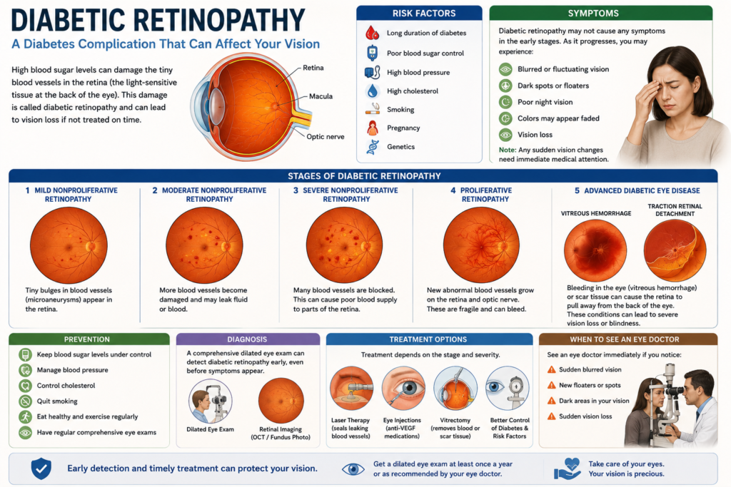
There’s a significant eye condition affecting individuals with diabetes called diabetic retinopathy. You are likely wondering about its origins and how it impacts your vision. Diabetic retinopathy is a complication of diabetes that damages blood vessels in the retina . It is a leading cause of irreversible blindness in working-age adults. Early detection and strict management of blood sugar, blood pressure, and cholesterol are vital to preventing vision loss. Diabetic retinopathy is most commonly referred to as diabetic eye disease . This post will explain what diabetic retinopathy is, its symptoms, and natural approaches to managing the condition.
Key Takeaways:
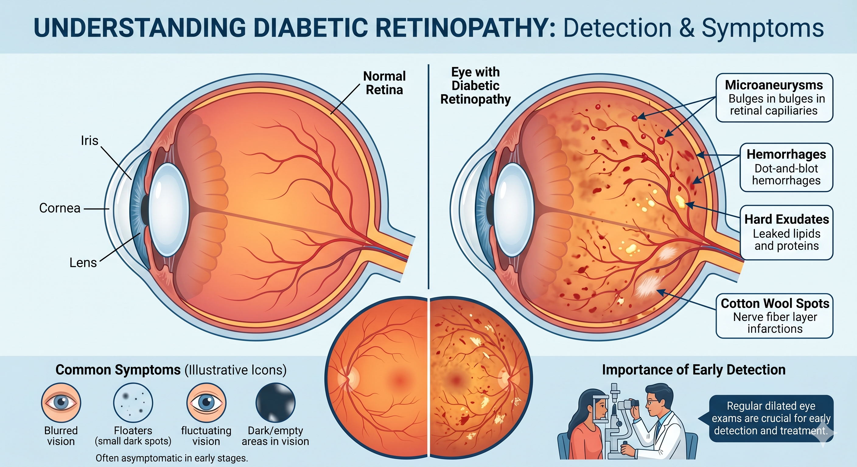
* Diabetic retinopathy, a complication of diabetes, damages blood vessels in the retina, potentially leading to vision loss. High blood sugar levels weaken these vessels over time, causing them to leak fluid or bleed.
* The condition progresses through stages, beginning with non-proliferative diabetic retinopathy (NPDR), where small blood vessel abnormalities occur. Advanced stages involve proliferative diabetic retinopathy (PDR), characterized by the growth of new, fragile blood vessels that can bleed and scar.
* Early stages of diabetic retinopathy often present without noticeable symptoms. Blurred vision, floaters, or dark spots typically appear as the condition advances, making regular eye exams crucial for early detection.
* Managing blood sugar, blood pressure, and cholesterol levels effectively slows the progression of diabetic retinopathy. This foundational control is a primary “natural treatment” approach.
* While no “natural cure” exists for advanced diabetic retinopathy, certain supplements, such as lutein, zeaxanthin, omega-3 fatty acids, and specific antioxidants, may support eye health and potentially reduce risk factors when combined with medical guidance.
* Emerging evidence suggests acupuncture might help manage some symptoms or improve blood flow in the eyes for certain individuals. Homeopathic remedies lack scientific validation for treating diabetic retinopathy and should not replace conventional medical care.
* Conventional treatments range from laser therapy (photocoagulation) to injections of anti-VEGF medications or steroids, and in advanced cases, vitrectomy surgery. These interventions aim to stop blood vessel leakage, reduce new vessel growth, and preserve vision.
Pathophysiology and Mechanisms of Retinal Damage
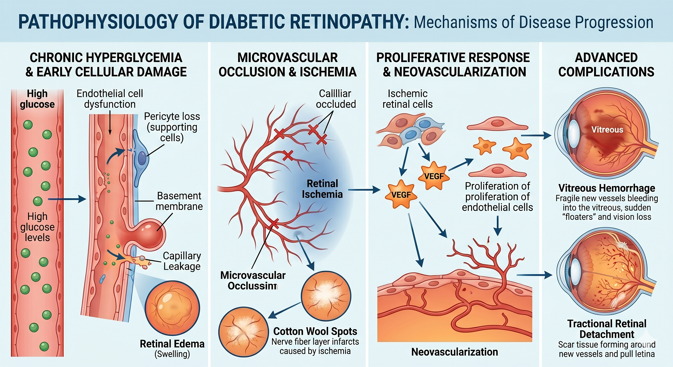
You might wonder how diabetes specifically damages your eyes. Diabetic retinopathy pathophysiology describes a complicated interaction of factors that compromise the delicate retinal environment. High blood sugar levels, a hallmark of diabetes, initiate a cascade of detrimental changes within the tiny blood vessels that nourish your retina. This chronic exposure to hyperglycemia sets the stage for the initial vascular alterations, ultimately leading to vision impairment. This process involves several key mechanisms, each contributing to the overall pathology. Oxidative stress, for instance, plays a significant role, as does the activation of various inflammatory pathways. These molecular changes culminate in the breakdown of the blood-retinal barrier, allowing fluid and proteins to leak into the retina. Understanding these underlying mechanisms is vital for appreciating the progressive nature of diabetic retinopathy. Ultimately, the sustained damage to these blood vessels can lead to their complete occlusion or the growth of abnormal, fragile new blood vessels. These new vessels are prone to bleeding, further exacerbating retinal damage and leading to severe vision loss. The progression from mild non-proliferative retinopathy to severe proliferative retinopathy is a direct consequence of these ongoing pathophysiological changes within your eye.
Vascular Changes in the Ocular Environment
The smallest blood vessels in your retina are particularly vulnerable to the effects of diabetes. These microvessels, vital for supplying oxygen and nutrients, undergo significant structural and functional changes. You will find that the endothelial cells lining these capillaries damage themselves, leading to increased permeability and leakage. Your retinal capillaries can also become blocked, depriving areas of the retina of vital blood flow. This ischemia triggers the body’s natural response to grow new blood vessels, a process known as neovascularization. However, these new vessels are often weak and abnormal, contributing to further complications. These vascular changes are a defining characteristic of diabetic retinopathy, directly impacting your visual acuity. The compromised blood-retinal barrier and the formation of leaky or occluded vessels are key to the development and progression of the disease in your eyes.
The Impact of Chronic Hyperglycemia on Retinal Health
Chronic high blood sugar, or hyperglycemia, directly instigates a series of biochemical reactions detrimental to your retinal cells. You experience increased oxidative stress, where an imbalance between free radicals and antioxidants damages cellular components. This stress contributes significantly to the breakdown of the blood-retinal barrier. Persistent hyperglycemia also heightens your body’s inflammatory responses. Various inflammatory mediators cause more damage to the delicate retinal tissues and contribute to leakage from blood vessels. This sustained inflammation perpetuates a cycle of damage within your eyes. Glycation end products, or AGEs, accumulate in your retinal tissues due to chronic hyperglycemia. These harmful compounds contribute to the stiffening and dysfunction of blood vessels, accelerating the progression of retinopathy. This accumulation of AGEs is a direct consequence of prolonged elevated blood sugar. The persistent elevation of glucose levels in your bloodstream triggers several harmful pathways that directly impact the delicate structures of your retina. For example, the polyol pathway becomes overactive, leading to the accumulation of sorbitol within retinal cells, which can cause osmotic stress and cellular damage. Additionally, the activation of protein kinase C (PKC) isoforms, which also happens because of hyperglycemia, leads to increased vascular permeability and abnormal growth of new blood vessels, making your retinal health worse.
Classification and Progression of Diabetic Eye Disease Stages
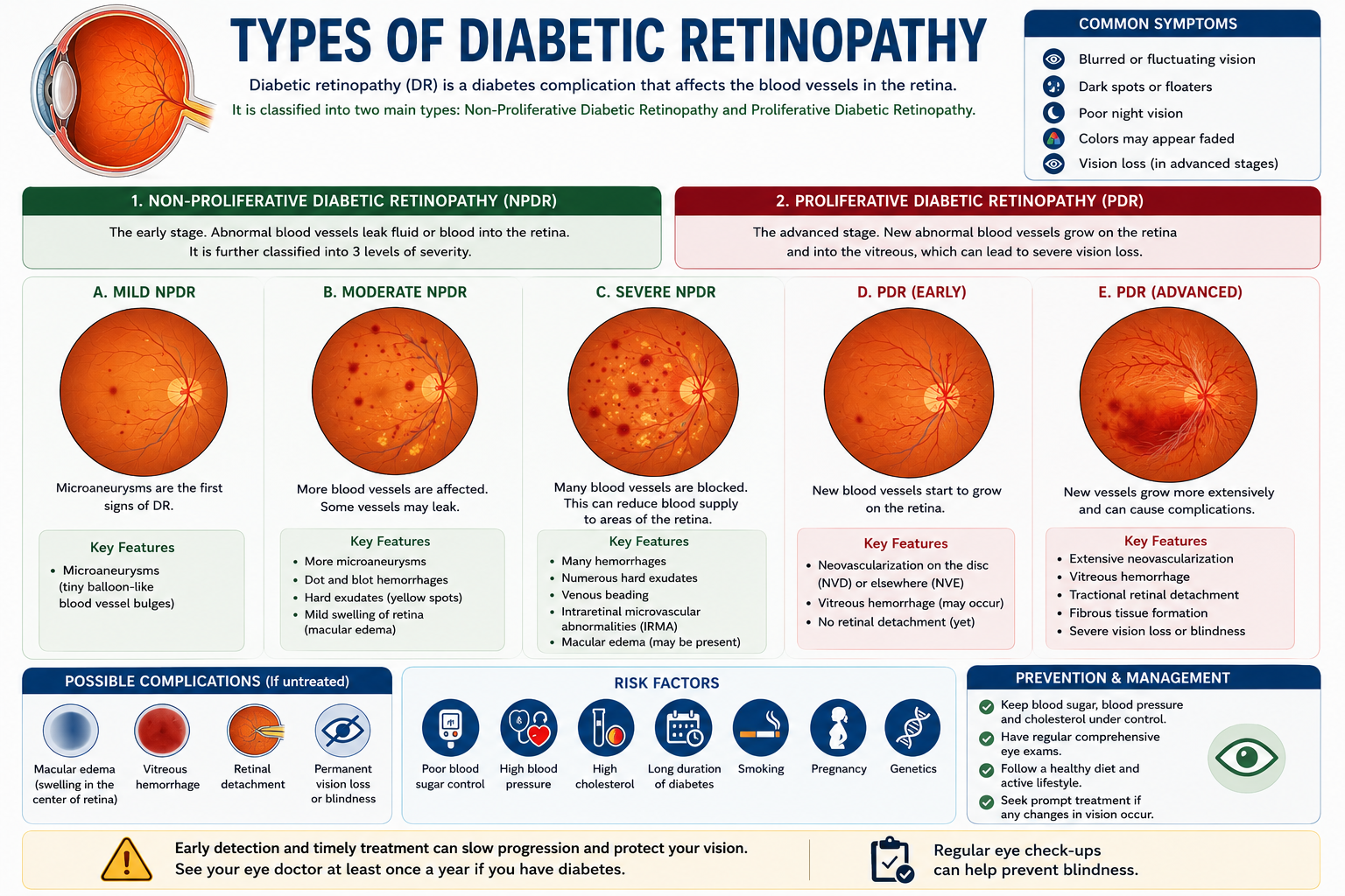
Understanding the progression of diabetic retinopathy is imperative for effective management and preservation of your vision. This condition unfolds in distinct stages, each characterized by specific changes within your retina. Recognizing these stages helps you and your healthcare provider anticipate potential complications and tailor interventions accordingly, emphasizing the importance of regular eye examinations. The journey of diabetic retinopathy typically encompasses four stages, evolving from mild to severe. These stages are broadly categorized into two main forms: non-proliferative diabetic retinopathy (NPDR) and proliferative diabetic retinopathy (PDR). The distinction between proliferative and non-proliferative diabetic retinopathy lies in the presence or absence of new, abnormal blood vessel growth, which indicates a significant change in the disease’s severity. You can think of these stages as a spectrum, with each step representing an increase in the damage to your retinal blood vessels. Early detection of any of these diabetic retinopathy stages allows for proactive measures to slow or even halt further advancement, protecting your precious eyesight from irreversible harm.
Characteristics of Non-Proliferative Diabetic Retinopathy Development
When your retina begins to show the initial signs of damage, you are in the non-proliferative diabetic retinopathy (NPDR) phase. This early stage is often asymptomatic, meaning you might not experience any noticeable changes in your vision. However, a comprehensive eye exam can reveal subtle but significant alterations within your retinal blood vessels. Tiny bulges, known as microaneurysms, are characteristic of mild NPDR. These small swellings in the blood vessel walls can leak fluid, causing the retina to swell. As the disease progresses to moderate NPDR, more microaneurysms appear, and small hemorrhages, or bleeding spots, become evident on your retina. Severe NPDR signifies a more extensive blockage of retinal blood vessels. This blockage deprives areas of your retina of important oxygen and nutrients, leading to a build-up of waste products. These changes serve as a warning sign, indicating that your eye is preparing for the more advanced, proliferative stage if left unaddressed.
Identifying Advanced Proliferative Changes
The shift to proliferative diabetic retinopathy (PDR) represents a critical escalation in the disease’s progression. This stage is defined by the growth of new, abnormal blood vessels on the surface of your retina and into the vitreous gel. These new vessels, known as neovascularization, are fragile and prone to bleeding. These delicate new blood vessels can easily rupture, leading to vitreous hemorrhage. Blood within the vitreous can significantly obscure your vision, sometimes causing sudden, severe vision loss. The body’s attempt to heal this bleeding can also result in scar tissue formation, which can pull on the retina. The scar tissue contraction can lead to retinal detachment, a serious condition where the retina pulls away from the back of the eye. Retinal detachment is a medical emergency that can cause permanent vision loss if not treated promptly. Understanding these advanced changes is crucial for seeking immediate medical attention. You might experience sudden floaters, flashes of light, or a dark curtain moving across your field of vision if you develop proliferative diabetic retinopathy. These symptoms indicate a serious problem that requires immediate evaluation by an ophthalmologist. Early intervention in PDR can prevent irreversible damage and preserve your sight.
- New, abnormal blood vessel growth (neovascularization)
- Vitreous hemorrhage from fragile vessels
- Formation of scar tissue on the retina
- Risk of retinal detachment
| Proliferative Diabetic Retinopathy Indicators | |
|---|---|
| Neovascularization | Abnormal blood vessel growth on the retina’s surface. |
| Vitreous Hemorrhage | Bleeding into the vitreous gel occurs due to fragile vessels. |
| Scar Tissue Formation | Fibrous tissue development that can pull on the retina. |
| Retinal Detachment Risk | Separation of the retina from the underlying tissue. |
Differential Diagnosis in Retinal Vascular Disease
Distinguishing between various retinal vascular diseases is paramount for accurate diagnosis and effective management. You will often encounter conditions presenting with similar symptoms, yet originating from entirely different systemic issues. A precise differential diagnosis ensures that you receive the most appropriate treatment, preventing unnecessary interventions and focusing on the underlying cause of your vision changes. Misdiagnosis can lead to ineffective therapies and continued progression of the disease, making a thorough evaluation by your ophthalmologist vital. Understanding the subtle differences in presentation allows for a targeted approach to your care. For instance, while both diabetic retinopathy and hypertensive retinopathy can cause vision loss, their specific retinal manifestations and progression patterns differ significantly. Your doctor will carefully examine your retina for characteristic signs, such as microaneurysms in diabetic retinopathy or arteriolar narrowing and cotton wool spots in hypertensive retinopathy, to determine the exact nature of your condition. This careful observation, combined with your medical history, forms the bedrock of a correct diagnosis. You play an active role in this diagnostic process by providing a comprehensive medical history, including any pre-existing conditions like diabetes or hypertension. This information, along with advanced imaging techniques like fundus photography and optical coherence tomography (OCT), helps your healthcare team accurately tell apart different retinal vascular diseases. A collaborative effort between you and your ophthalmologist ensures that the diagnostic journey is thorough, leading to the most effective treatment strategy for preserving your vision.
Comparative Analysis of Vascular Retinopathies
You may find it challenging to discern the subtle differences between various vascular retinopathies, but your ophthalmologist is trained to identify key distinguishing features. While both hypertensive retinopathy and diabetic retinopathy affect the retinal vasculature, their underlying pathogenic mechanisms and characteristic retinal signs differ significantly. Recognizing these distinctions is crucial for accurate diagnosis and guiding appropriate treatment strategies for your specific condition.
Consider the primary drivers behind each condition: diabetic retinopathy stems from prolonged hyperglycemia causing damage to the small blood vessels of the retina, leading to microaneurysms, hemorrhages, and eventually neovascularization. Hypertensive retinopathy, on the other hand, results from chronic high blood pressure damaging the retinal arterioles, causing narrowing, exudates, and cotton wool spots. You will notice that the initial changes in each condition often manifest in distinct ways, providing valuable clues for your doctor.
The progression of these diseases also follows different trajectories. Diabetic retinopathy can advance through non-proliferative stages to proliferative diabetic retinopathy, characterized by new, fragile blood vessel growth that can lead to severe vision loss. Hypertensive retinopathy’s severity is often directly correlated with the degree and duration of uncontrolled hypertension, with signs often improving with blood pressure control. Understanding these divergent paths helps your doctor predict the course of your disease and tailor your management plan effectively.
Hypertensive Retinopathy vs. Diabetic Retinopathy
While both diabetic retinopathy (DR) and hypertensive retinopathy (HR) are vascular diseases that damage the blood vessels of the retina, they have different underlying causes, mechanisms, and hallmark visual signs. Because patients often have both diabetes and hypertension simultaneously, distinguishing between the two is a critical component of a comprehensive eye exam.
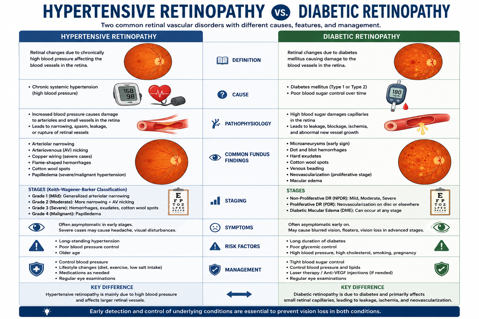
Here is a breakdown of the differential diagnosis between the two conditions.
Quick Comparison
| Feature | Diabetic Retinopathy (DR) | Hypertensive Retinopathy (HR) |
| Primary Cause | Chronic hyperglycemia (high blood sugar) | Chronic or acute elevated blood pressure |
| Pathophysiology | Capillary leakage, microvascular occlusion, and ischemia leading to neovascularization. | Arteriospasm, thickening of vessel walls (arteriolosclerosis), and breakdown of the blood-retinal barrier. |
| Typical Hemorrhages | Dot-and-blot hemorrhages (deep in the retina). | Flame-shaped hemorrhages (superficial nerve fiber layer). |
| Vascular Changes | Microaneurysms are the earliest clinical sign, and venous beading is also present. | Arteriolar narrowing, AV nicking, and copper/silver wiring. |
| Exudate Pattern | Hard exudates (lipid deposits), often scattered or in circinate (ring) patterns. | Hard exudates can classically form a macular star in severe cases. |
| Advanced Complications | Proliferative neovascularization, vitreous hemorrhage, and tractional retinal detachment. | Optic disc edema (papilledema), typically seen in malignant hypertension. |
Hallmarks of Diabetic Retinopathy (DR)
Diabetic retinopathy primarily affects the venous and capillary systems of the retina. The chronic high blood sugar damages the endothelial cells and pericytes (supporting cells) of the capillaries, causing them to leak or become blocked.
Microaneurysms: Small, red dots on the retina. These are often the very first clinical signs of DR and represent localized outpouchings of damaged capillaries.
Dot-and-Blot Hemorrhages: Bleeding that occurs in the deeper layers of the retina, taking on a round, well-defined appearance.
Venous Beading: Retinal veins that develop irregular, sausage-like segments due to sluggish blood flow and ischemia.
Neovascularization: In the advanced “proliferative” stage, the ischemic retina releases Vascular Endothelial Growth Factor (VEGF), prompting the growth of fragile, abnormal new blood vessels that easily bleed into the vitreous.
Hallmarks of Hypertensive Retinopathy (HR)
Hypertensive retinopathy primarily affects the arterial system of the retina. High blood pressure forces the vessels to adapt by thickening their walls, which ultimately restricts blood flow and damages the vessels.
AV Nicking (Arteriovenous Crossing Changes): Because retinal arteries and veins share a common adventitial sheath where they cross, a thickened, hardened artery will compress the underlying vein, making the vein look “pinched” or “nicked.”
Arteriolar Narrowing & Wiring: The arteries become narrowed and their reflective properties change. They may look like copper wires (copper wiring) or, in more advanced sclerosis, silver wires (silver wiring).
Flame-Shaped Hemorrhages: Bleeding in the superficial nerve fiber layer of the retina, which follows the architecture of the nerve fibers, giving them a splintered or flame-like appearance.
Macular Star and Papilledema: In severely high, uncontrolled blood pressure (malignant hypertension), fluid leaks into the macula, forming a star-like pattern of dense exudates, and the optic nerve head swells (papilledema).
Areas of Overlap
It is important to note that because both conditions cause ischemia (lack of oxygen) and damage to the blood-retinal barrier, they share a few non-specific findings:
Cotton Wool Spots: These fluffy white patches are areas of nerve fiber layer infarction (localized cell death due to lack of blood supply) and can appear in both conditions.
Macular Edema: Swelling in the central area of the retina due to fluid leakage can be caused by both DR and HR, though it is a more prominent and common cause of vision loss in diabetes.
Holistic and Natural Treatment Paradigms
Recognizing the complex nature of diabetic retinopathy reveals the importance of a comprehensive strategy that extends beyond conventional approaches. You might be surprised to learn that many individuals are actively exploring natural treatments for diabetic retinopathy, aiming not just to manage but potentially reverse the condition. This paradigm shift acknowledges the body’s inherent healing capabilities when supported by the right nutritional and lifestyle interventions. The goal is to address the root causes of retinal damage, moving beyond symptom management to true cellular repair and regeneration. Exploring natural treatment for diabetic retinopathy involves a multi-faceted approach, often integrating dietary changes, specific supplements, and lifestyle modifications. You’ll find that many of these strategies focus on reducing inflammation, improving blood circulation, and strengthening blood vessel integrity, all of which are critical for eye health. The concept of reversing diabetic retinopathy naturally resonates with those seeking alternatives to more invasive procedures, offering hope for regaining visual acuity and preventing further progression. Considering the potential for reversing diabetic retinopathy naturally, it’s necessary to approach this topic with informed choices and guidance. You can research various natural treatments for diabetic retinopathy, often encountering success stories that highlight the power of these integrated methods. This section will guide you through both conventional management and the exciting possibilities of integrated natural strategies, providing a balanced perspective on how you can actively participate in your eye health journey.
Conventional Management and Therapeutic Goals
Traditional diabetic retinopathy treatment primarily focuses on halting the progression of the disease and preserving existing vision. You’ll typically encounter options like laser photocoagulation, which aims to seal off leaky blood vessels or destroy abnormal ones, and intravitreal injections of anti-VEGF medications, designed to reduce swelling and abnormal blood vessel growth. These treatments are often initiated when the retinopathy reaches a certain severity, such as proliferative diabetic retinopathy or diabetic macular edema. Your ophthalmologist will work to stabilize your condition and prevent further vision loss, which is the primary therapeutic goal of conventional diabetic retinopathy treatment. Regular eye examinations are crucial to monitor the progression of the disease and determine the optimal timing for interventions. The aim is to mitigate the immediate threats to your vision, acting as a critical line of defense against severe and irreversible damage caused by uncontrolled diabetes. These conventional methods do not typically address the underlying metabolic imbalances that contribute to diabetic retinopathy, even though they are effective in many cases for managing immediate symptoms. You might find that while these treatments can slow down the disease, they don’t necessarily provide a pathway for reversing diabetic retinopathy naturally. This distinction is important when considering a holistic approach to your eye health.
Integrated Strategies for Natural Reversal
You can actively pursue reversing diabetic retinopathy naturally by adopting a comprehensive integrated strategy. This approach focuses on optimizing your overall health, recognizing that the eyes are a reflection of systemic well-being. Many natural treatments for diabetic retinopathy protocols emphasize rigorous blood sugar control, which is undeniably the cornerstone of preventing and potentially reversing diabetic complications. Implementing specific dietary changes is a key component of reversing diabetic retinopathy naturally. You should consider a diet rich in antioxidants, vitamins, and minerals, focusing on whole, unprocessed foods. Eliminating inflammatory foods and incorporating those known to support eye health, like leafy greens, berries, and omega-3-rich foods, can significantly contribute to natural treatment for diabetic retinopathy. Supplementation with specific nutrients also plays an important role in natural treatment for diabetic retinopathy. You might explore supplements such as alpha-lipoic acid, lutein, zeaxanthin, and bilberry extract, all of which have been studied for their potential benefits in supporting retinal health and improving microcirculation. These integrated strategies work synergistically to create an environment conducive to healing and repair within the delicate structures of your eyes. You can further enhance your integrated strategy by incorporating regular exercise, stress reduction techniques, and ensuring adequate sleep, all of which contribute to better blood sugar management and overall cellular health. These lifestyle changes, along with specific nutritional interventions, form a strong approach to naturally reverse diabetic retinopathy, providing a proactive and empowering way to improve vision and eye health.
Complementary Modalities and Nutritional Support for Diabetic Retinopathy
You might be exploring additional avenues to support your retinal health beyond conventional treatments. While medical interventions are crucial for managing diabetic retinopathy, a holistic approach often includes complementary modalities and targeted nutritional strategies. These methods aim to reduce inflammation, improve circulation, and protect delicate retinal cells from further damage, working in conjunction with your doctor’s recommendations. Consider the powerful role that specific nutrients and alternative therapies can play in your overall management plan. Many individuals find that a combination of approaches helps to mitigate symptoms and potentially slow the progression of diabetic retinopathy. You are actively taking steps to protect your vision by investigating these supportive measures. Integrating these complementary strategies into your daily routine can offer a sense of proactive engagement in your health journey. Always discuss any new supplements or therapies with your healthcare provider to ensure they are safe and appropriate for your individual circumstances. Your well-being is the top priority in this comprehensive management strategy.
Targeted Supplementation for Retinal Integrity
Specific supplements can offer significant support for the delicate structures of your retina. Antioxidants, for instance, play a critical role in neutralizing free radicals that contribute to cellular damage in diabetic retinopathy. You might consider compounds like lutein and zeaxanthin, known for their protective effects on the macula, filtering harmful blue light and reducing oxidative stress. Certain vitamins and minerals are also vital for maintaining healthy blood vessels and nerve function in the eye. Alpha-lipoic acid, another potent antioxidant, has shown promise in supporting nerve health and improving blood sugar control. You may also find benefits from omega-3 fatty acids, particularly DHA, which is a major structural component of retinal cell membranes and possesses anti-inflammatory properties. Many individuals explore a comprehensive approach that includes a blend of these supportive nutrients. Resveratrol, found in red grapes, is another compound with antioxidant and anti-inflammatory effects that could contribute to retinal health. You are investing in your long-term vision by providing your body with these targeted nutritional building blocks.
Alternative Medicine and Acupuncture Applications for Diabetic Retinopathy
Exploring alternative medicine offers additional pathways to support your retinal health. Acupuncture, an ancient practice, has been studied for its potential to improve blood flow to the eyes and reduce inflammation. You might find that sessions with a qualified acupuncturist help alleviate some of the discomfort associated with diabetic retinopathy and promote overall ocular well-being. Homeopathic medicine presents another avenue for individuals seeking natural and individualized treatment approaches. This system focuses on stimulating the body’s self-healing mechanisms through highly diluted substances. You would work with a homeopathic practitioner to identify remedies specifically tailored to your unique symptoms and constitution, aiming to address the root causes of the imbalance. These alternative therapies can be valuable additions to your existing treatment plan, providing complementary support. You are empowering yourself by exploring a wider range of options that can contribute to your visual health and overall quality of life. Acupuncture, specifically, involves the insertion of thin needles into specific points on the body, aiming to balance energy flow and stimulate healing. Studies have indicated that acupuncture can improve microcirculation, which is crucial for delivering oxygen and nutrients to the retina. For individuals with diabetic retinopathy, this improved blood flow could potentially help in reducing retinal ischemia and supporting the health of the existing blood vessels.
Acupuncture for Diabetic Retinopathy
- Potential Benefits : Recent systemic reviews indicate that acupuncture—especially when combined with traditional medications—may improve visual acuity and reduce central macular thickness.
- Mechanisms : Clinical studies suggest it may enhance blood microcirculation behind the eye, reduce retinal capillary lesions, and alleviate systemic inflammation.
- Limitations : The current body of scientific evidence carries a high risk of bias, small sample sizes, and inconsistent methodologies. No definitive, large-scale clinical trials have proven it can halt disease progression on its own.
- Needle Placement : Certified practitioners insert fine needles into specific acupoints on the hands, feet, ears, and eyebrows.
- Safety Restraint : No needles are ever placed directly into the eyeball .
- Electroacupuncture : Some clinics pass a mild, low-intensity electrical current through the needles to further stimulate nerve pathways and local blood flow.
Homeopathic Management of Diabetic Retinopathy
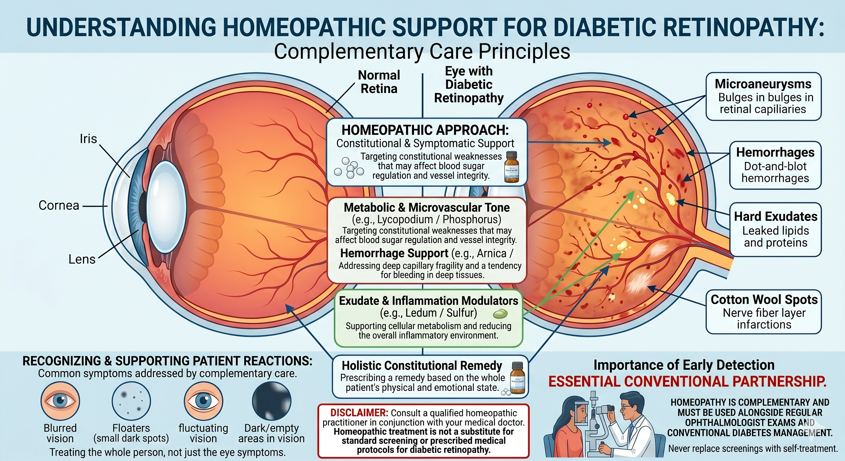
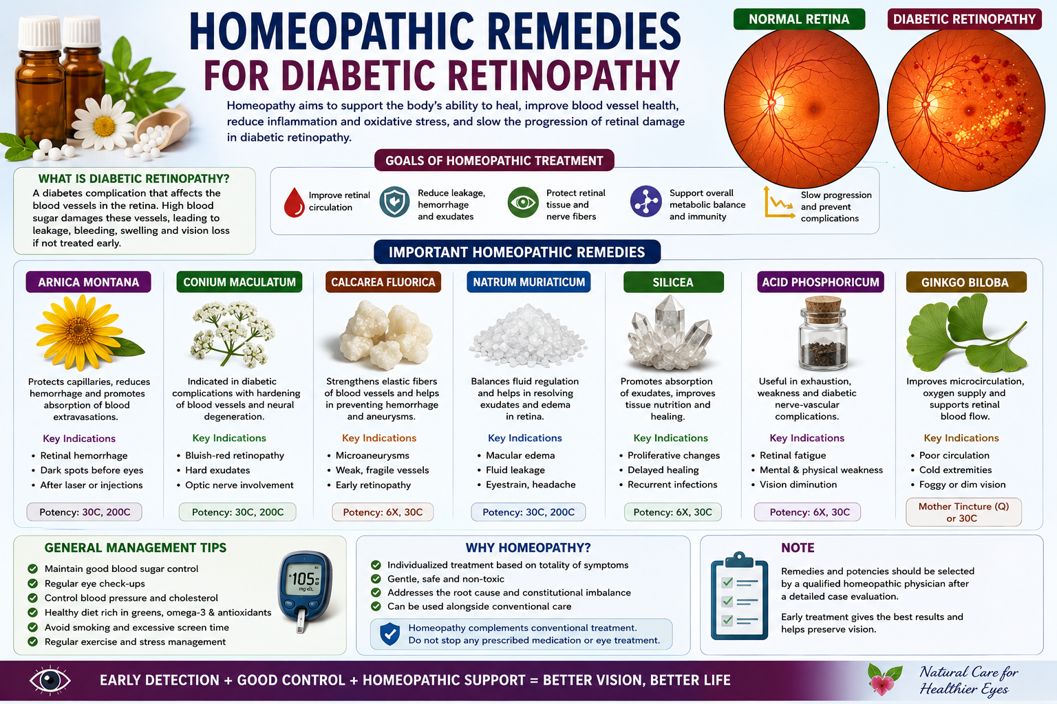
Homeopathic treatment for diabetic retinopathy offers several potential benefits, which can complement conventional medical approaches. Homeopathy works with the body’s natural healing processes, stimulating the immune system and enhancing the body’s ability to heal itself. By addressing the root cause of the condition, rather than just masking the symptoms, homeopathic treatment aims to promote long-term health and vitality. Homoeopathic medicine has a role to play in various stages of the ailment. It has remedies to improve the perfusion of the retina with Lithium carb, Nux vomica, and Ginkgo biloba. When changes are first observed, phosphorus is one of the remedies that can help prevent the progression of retinopathy in the blood vessels.
Homeopathic remedies
Phosphorus
Phosphorus symptoms include black patches in front of the eyes, light sensitivity, blurred vision, and retinal bleeding. Sensitive, nervous, and lonely are all examples of personality traits. Beneficial in: Diabetic Retinopathy with floaters or beginning bleeding.
Jambolanum Syzygium
Indications: Well-known for naturally regulating blood sugar levels. Use: Although not eye-specific, it is used as a supportive treatment for diabetes, which inadvertently preserves normal eye health in spite of the disease.
Montana Arnica
Indications include retinal hemorrhage, trauma-related injury, and eye strain. Beneficial in: Diabetic retinopathy with bleeding or bruises in the eyes. “As if the eyes have been beaten” is the keynote.
Horridus Crotalus
Suggestions: Tendency to bleed, especially when there is retinal hemorrhage. Beneficial in: Severe clotting or recurrent bleeding instances.
Lycopodium
Suggestion: DME (Macular Oedema), with dim or blurry vision, and digestive
FAQs

Can diabetic eye problems be treated with homeopathy?
Homeopathy can, in fact,
- slows down the development, especially in the early stages.
- can enhance blood flow to the retina.
- can strengthen blood vessels
- reduces associated symptoms like eye strain, floaters, and blurred vision.
It functions best when combined with routine eye and diabetes treatment.
Is homeopathic medicine safe for people with diabetes?
When used correctly, homeopathy is harmless and has no known adverse effects. It doesn’t interfere with insulin, ocular injections, or diabetes medications. However, a qualified homeopathic physician must always be consulted before taking it.
How long does it take for homeopathy to show results?
The answer differs depending on:
- The disease’s stage
- Your overall diabetes management
- The frequency and intensity of ocular symptoms
- People may see improvement in the early phases in a few weeks or months. It is primarily palliative in severe phases.
Can blood sugar be lowered by homeopathy?
Yes, when used in conjunction with diet, lifestyle changes, and allopathic treatment, medications like Syzygium Jambolanum Q and Cephalandra indica Q can help regulate blood sugar levels.
If I have diabetes, how often should my eyes be examined?
Answer: Even if you feel healthy, you should have a dilated eye exam every year. Your eye care specialist may advise you to visit more frequently if you already have an eye illness.
Is Diabetic Eye Disease Deadly?
No, diabetic eye disease is not fatal on its own.
Although it is not life-threatening, it is a significant complication of diabetes that, if left untreated, can result in lifelong blindness.
Conclusion
Presently, you understand diabetic retinopathy stems from prolonged high blood sugar damaging the retina’s blood vessels. This damage moves from non-proliferative diabetic retinopathy (NPDR), where microaneurysms and hemorrhages occur, to proliferative diabetic retinopathy (PDR), which is marked by neovascularization and may lead to retinal detachment. Recognizing symptoms like blurred vision, floaters, and vision loss early is paramount, as early detection allows for more effective intervention. Your proactive management of blood glucose levels, blood pressure, and cholesterol significantly mitigates the risk and progression of this condition. You can also explore natural treatment modalities, which complement conventional medical approaches. For instance, studies have indicated that saffron (Crocus sativus) may protect retinal cells from oxidative stress. Turmeric, specifically its active compound curcumin, exhibits anti-inflammatory properties that can benefit retinal health. Omega-3 fatty acids, found in fish oil, are known for their anti-inflammatory effects and their role in maintaining overall eye health. You should discuss these options with your healthcare provider to ensure they integrate safely and effectively with your existing treatment plan. Your ongoing commitment to a healthy lifestyle, including a balanced diet rich in antioxidants, regular exercise, and consistent monitoring of your blood sugar, remains your most powerful defense against diabetic retinopathy. Regular eye examinations with an ophthalmologist are indispensable for early diagnosis and timely intervention. By combining diligent self-care with informed natural approaches and professional medical guidance, you can actively preserve your vision and maintain your quality of life.
FAQ
Q: What is diabetic retinopathy?
A: Diabetic retinopathy is a complication of diabetes that affects the eyes. High blood sugar levels damage the blood vessels in the retina, the light-sensitive tissue at the back of the eye. This damage can lead to vision impairment and, if left untreated, blindness. The condition often progresses without early symptoms, making regular eye exams for individuals with diabetes critical.
Q: How does diabetes cause retinopathy?
A: Diabetes causes retinopathy through prolonged exposure to elevated blood glucose levels. These high sugar levels harm the tiny blood vessels supplying the retina. The walls of these vessels can weaken, bulge, and leak fluid or blood into the retina. New, abnormal blood vessels may also grow on the surface of the retina; these are fragile and prone to bleeding. This process disrupts the retina’s ability to send clear images to the brain, leading to blurry vision or dark spots.
Q: What are the four stages of diabetic retinopathy?
A: Diabetic retinopathy progresses through distinct stages. The first stage is mild non-proliferative diabetic retinopathy (NPDR), which is marked by microaneurysms, small bulges in the retinal blood vessels. Moderate NPDR shows more extensive microaneurysms, hemorrhages, and cotton wool spots, which are areas of nerve fiber damage. Severe NPDR involves numerous hemorrhages and blockages in blood vessels, signaling a high risk for the next stage. Proliferative diabetic retinopathy (PDR) is the advanced stage where new, abnormal blood vessels grow on the retina’s surface. These new vessels are fragile and can bleed into the vitreous gel, causing significant vision loss. Scar tissue can also form, pulling on the retina and potentially leading to retinal detachment.
Q: What is the first sign of diabetic retinopathy?
A: Many people with diabetic retinopathy experience no symptoms in its early stages. The first noticeable sign can be blurry vision, especially when reading, or difficulty seeing at night. Some individuals may observe floaters, which are specks or cobweb-like images that drift across their field of vision. A sudden increase in floaters or flashes of light could indicate bleeding in the eye or a serious retinal issue. Vision loss, dark spots, or a sudden, dramatic decrease in vision are signs of advanced disease requiring immediate medical attention.
Q: What are some natural approaches that might support eye health for individuals with diabetic retinopathy?
A: While conventional medical treatments are primary for diabetic retinopathy, certain lifestyle and dietary adjustments can support overall eye health. Strict blood sugar control, achieved through diet, exercise, and medication, is the most effective natural step. A diet rich in antioxidants, found in fruits, vegetables, and leafy greens, can help protect eye cells from damage. Omega-3 fatty acids, present in fish oil, may also have protective effects on the retina. Regular physical activity improves circulation and helps manage blood sugar. Some individuals explore supplements like lutein, zeaxanthin, and vitamins C and E, but scientific evidence directly reversing retinopathy with these is limited. Always discuss any natural treatments or supplements with your doctor to ensure they are safe and do not interfere with prescribed medications.
Q: Can acupuncture or homeopathic medicine treat diabetic retinopathy?
A: Acupuncture and homeopathic medicine are alternative therapies, and there is limited scientific evidence to support their effectiveness in treating or reversing diabetic retinopathy. Acupuncture involves inserting thin needles into specific points on the body. Some proponents suggest it may improve blood flow or reduce inflammation, but its direct impact on retinal vessel damage in diabetic retinopathy is not established by rigorous studies. Homeopathic medicine uses highly diluted substances based on the principle of “like cures like.” No scientific research has demonstrated that homeopathic remedies can prevent, treat, or reverse the progression of diabetic retinopathy. Individuals considering these therapies should understand they are not substitutes for conventional medical care and should always consult their ophthalmologist before trying any alternative treatments.
Q: What are the main differences between hypertensive retinopathy and diabetic retinopathy?
A: Both hypertensive retinopathy and diabetic retinopathy affect the retinal blood vessels, but their underlying causes and specific manifestations differ. Hypertensive retinopathy results from chronically high blood pressure, which causes hardening and narrowing of the retinal arteries. This can lead to retinal hemorrhages, exudates (leaky fluid), and optic nerve swelling in severe cases. Diabetic retinopathy, as discussed, is caused by high blood sugar damaging retinal vessels, leading to microaneurysms, leakage, and abnormal vessel growth. While both conditions can cause vision loss, the specific types of vessel damage and the progression patterns are distinct. An ophthalmologist can differentiate between the two conditions through a comprehensive eye exam, as treatment approaches vary based on the underlying cause.
Natural Treatment for Diabetic Retinopathy in Philadelphia
If you are living in the City of Brotherly Love and suffer from diabetic retinopathy, contact Philadelphia Holistic Clinic to schedule an appointment with Victor Tsan.
At the clinic, you will receive a comprehensive holistic evaluation and customized natural treatment. All holistic modalities, including acupuncture, homeopathy, reiki, psychotherapy, etc., are available at the Philadelphia Holistic Clinic.