Dyshidrotic Eczema
Small, fluid-filled blisters erupting along the edges of your fingers or across your palms can transform a simple handshake into a moment of sharp discomfort. This hallmark presentation often points to dyshidrotic eczema, a specific form of dermatitis also identified medically as dyshidrosis. Unlike surface-level dryness, the condition drives an intense, deep-seated itch beneath the skin, typically followed by a prickling or burning sensation long before any visible marks appear. Defined by recurrent flare-ups that cycle unpredictably, dyshidrosis sets itself apart from other eczemas through the unique location and nature of its eruptions, which may persist for several weeks before the skin begins to peel and crack open.
An outbreak does not discriminate by age, though adults between 20 and 40 years old report the highest incidence. Women and individuals who have a personal or family history of atopic dermatitis, hay fever, or asthma are more likely to experience the condition the condition surfaces more readily. Warm, humid weather acts as a frequent catalyst, causing sweat glands to become trapped beneath a compromised skin barrier, yet winter’s dry indoor heating can be just as provocative. Occupations requiring frequent hand washing or prolonged contact with metal salts—seen in mechanics, healthcare workers, and hair stylists—correlate with a greater likelihood of developing the characteristic vesicles on the lateral aspects of the digits.
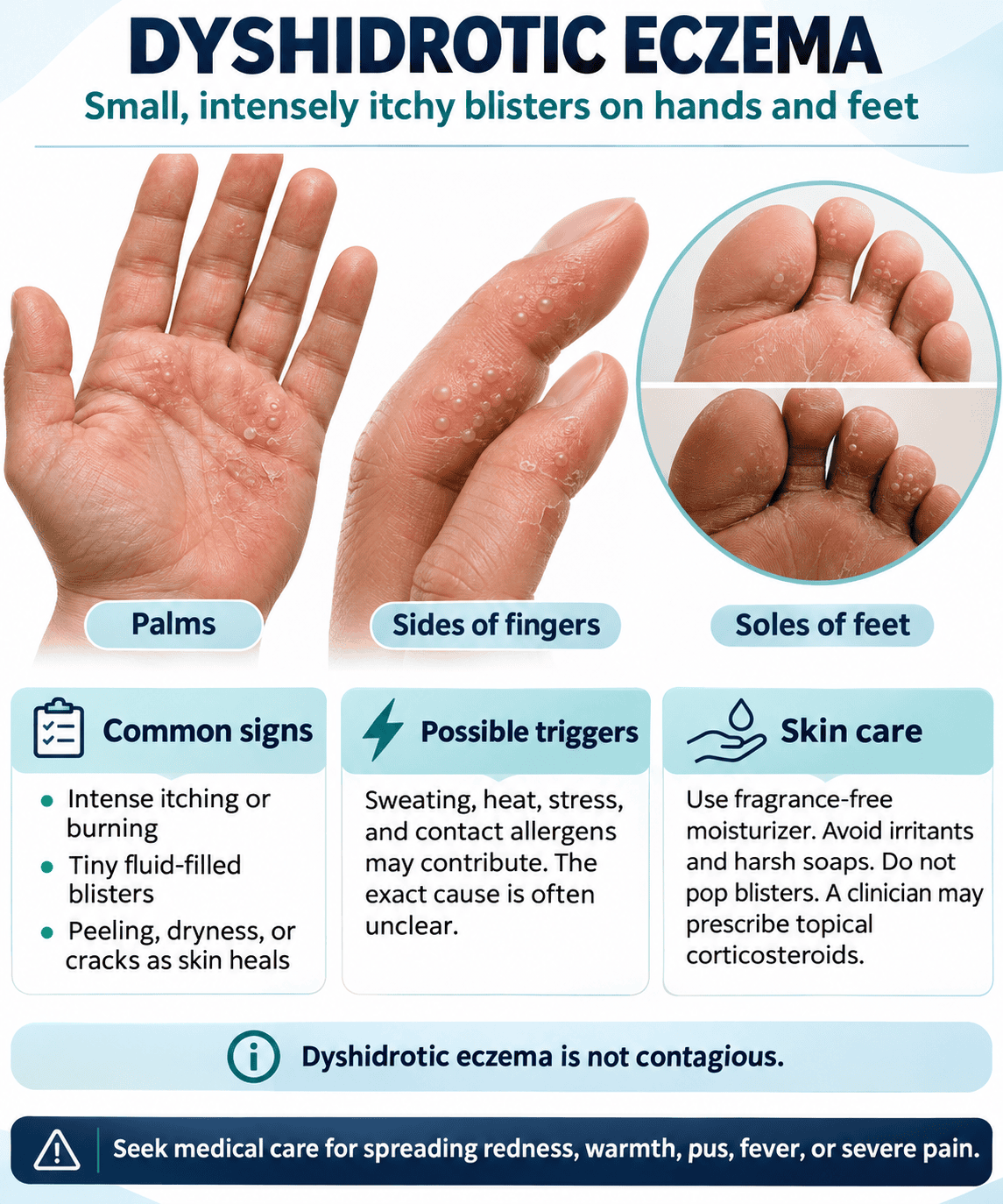
Recognizing dyshidrosis requires distinguishing it from contact dermatitis or fungal infections that share a similar visual footprint. The condition may resolve on its own without intervention, but the blistering phase often gives way to painful fissures that compromise your skin’s protective function for days afterward. Nicknamed “housewife’s eczema” in older medical literature due to its association with water exposure, the terminology has since evolved to reflect a broader understanding of genetic susceptibility and environmental interplay. Managing this cycle involves more than suppressing a rash; it calls for an approach that addresses skin barrier repair alongside the identification of hidden dietary or contact irritants that ignite the immune response.
Key Takeaways:
- The condition manifests as clusters of deep, tapioca-textured blisters on the palms, sides of fingers, and soles, and is distinguished from other forms of dermatitis by the absence of redness and scale in the very earliest stages.
- A disrupted skin barrier function, often linked to a filaggrin gene mutation, serves as the foundational vulnerability that allows common triggers like nickel, cobalt, and prolonged moisture to initiate a flare, explaining why some people react while others do not.
- Nickel and cobalt in the diet, found in foods such as whole grains, legumes, and chocolate, are now recognized as systemic triggers that can provoke a flare without any direct skin contact with metal, which shifts the focus from purely external causes to internal management.
- Psychological stress is not merely a secondary aggravator but can act as a primary trigger by prompting the release of histamine and inflammatory neuropeptides from nerve endings in the skin, a phenomenon that often explains recurring flares during otherwise well-managed periods.
- Diluted bleach baths, using a concentration of roughly 0.005% sodium hypochlorite similar to a chlorinated swimming pool, offer a low-cost method to reduce staphylococcal colonization on the affected skin, which in turn diminishes secondary infection and the inflammatory stimulus it provides.
- Moisturizer selection should prioritize products with a high lipid-to-water ratio, such as ointments or balms containing ceramides and urea, as these repair the intercellular lipid matrix more effectively than lotions and directly address the defective barrier at the core of the pathology.
- A comprehensive holistic strategy pairs conventional short-term corticosteroid use for acute vesiculation with longer-term lifestyle modifications like wearing cotton-lined gloves during wet work, performing patch testing for contact allergens, and following a personalized low-nickel diet guided by a registered professional.
Causes and Triggers of Dyshidrotic Eczema
A definitive single origin for dyshidrotic eczema remains elusive, yet the medical consensus points toward a malfunction within the skin’s barrier function combined with an overactive immune response. Your sweat glands were once suspected as the primary culprit, but research has since disproven this, showing the blisters form because the outermost layer of your epidermis, the stratum corneum, breaks down and allows irritants to penetrate far deeper than normal. This breakdown is often linked to a specific genetic filaggrin deficiency, a protein essential for maintaining a sturdy skin barrier, which leaves your pores vulnerable to inflammation that manifests as the deep, tapioca-like vesicles characteristic of the condition.
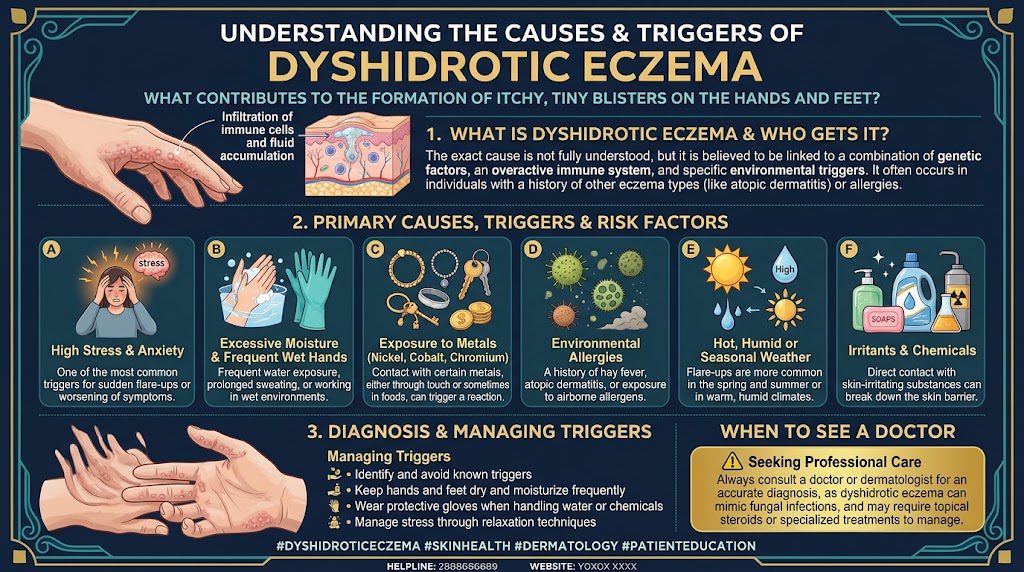
Atopic predisposition acts as the most significant internal driver for this disorder. Nearly 50 percent of individuals who develop the hallmark blisters on their palms, fingers, or soles have a personal or family history of atopic dermatitis, hay fever, or allergic asthma. Your immune system, primed by this atopic background, generates an exaggerated immunoglobulin E (IgE) response upon contact with otherwise benign substances. Excess emotional stress further destabilizes the skin by releasing cortisol and catecholamines, a biological cascade that directly degrades the barrier function, explaining why many first outbreaks coincide with periods of high psychological strain rather than a change in physical exposures.
Systemic factors also play a critical, often underestimated role in initiating a flare. A dermatologist consult frequently looks beyond the skin to rule out an ID reaction, where a fungal infection such as tinea pedis in a distant location triggers a sterile blistering outbreak on the hands. Intravenous immunoglobulin therapy, prescribed for autoimmune conditions, causes dyshidrotic eruptions in a documented subset of patients, as do excessive cobalt and nickel concentrations sourced from dietary intake. A patient consuming large quantities of oats, nuts, and dark chocolate might unknowingly sustain a high nickel load, keeping the histamine-mediated response perpetually active without direct topical contact.
| Trigger Category | Specific Irritant or Allergen | Exposure Example |
|---|---|---|
| Contact Allergens | Nickel sulfate | Cell phones, zippers, keys, jewelry |
| Contact Allergens | Cobalt chloride | Pigments in paints, metal tools, certain ceramics |
| Contact Irritants | Methylisothiazolinone | Wet wipes, liquid soaps, household cleaners |
| Physical Factors | Hyperhidrosis (excess sweat) | Prolonged wearing of occlusive gloves or synthetic socks |
| Systemic Allergens | Dietary nickel | Whole grains, legumes, canned foods, shellfish |
Causes of dyshidrosis
A precise biological pathway activates the moment your skin’s structural integrity is compromised, permitting water loss through the epidermis. Specialized filaggrin proteins break down into natural moisturizing factor molecules, and a deficiency here diminishes the hydration and acidity of the stratum corneum, essentially removing the protective acid mantle. Without this slightly acidic pH, barrier lipids dissolve, and the keratinocytes physically shrink, creating microscopic cracks that let common haptens like nickel ions bind to skin proteins. Your Langerhans cells then present these newly formed antigens to T-lymphocytes, initiating the spongiotic tissue reaction visible under a microscope, characterized by intercellular edema and fluid-filled intraepidermal vesicles.
Linking the dyshidrotic eczema exclusively to environmental contact underestimates the endogenous hypersensitivity that dictates lesion distribution. The dense concentration of eccrine sweat glands on the palms and soles creates a unique osmotic environment where metal salts dissolved in sweat can penetrate the acrosyringium, the terminal duct of the sweat gland. A patch test confirming a nickel allergy does not fully explain why you might experience a symmetrical eruption on both hands after eating a high-cobalt meal, a phenomenon termed systemic allergic dermatitis. Internal metal loads bypass the skin surface entirely, circulating in the bloodstream to reach the hyper-vascularized palmoplantar tissue, depositing directly into the dermis and triggering a delayed type IV hypersensitivity response without physical handling of metal.
Pharmacological and physiological stresses push a genetically susceptible system past the threshold of tolerance. The rapid withdrawal of potent topical corticosteroids can induce a severe status cosmeticus, a rebound effect where an abrupt discontinuation of clobetasol propionate causes an eruption far more violent than the original dermatitis. Smoking exerts a potent oxidative stress on endothelial cells, doubling the likelihood of chronic hand involvement due to capillary constriction and the release of matrix metalloproteinases that degrade collagen support. A clinical audit of patients with recalcitrant dyshidrosis often uncovers a hidden tinea infection creating circulating antigens, a type I hypersensitivity to dermatophyte toxins that requires eradicating the foot fungus with terbinafine before the sterile hand blisters ever subside.
Common triggers of dyshidrotic eczema
Occupational moisture stands as the most pervasive and preventable trigger for ongoing dyshidrotic flares. Wearing occlusive rubber or nitrile gloves for over two hours without a cotton liner creates a macerated environment where the stratum corneum swells to three times its resting thickness and becomes permeable. Hairdressers exposing wet hands to ammonium persulfate bleach and hairdresser’s solution have a documented incidence rate of hand dermatitis reaching 38 percent within the first three years of their career. A mechanic repeatedly washing engine grime with abrasive industrial solvents strips the intercellular lipids that form the only defense against the nickel present in every tool handle they grip, creating the perfect storm for a bilateral palmar outbreak.
Seasonal pollen surges and ultraviolet radiation introduce a less recognized but potent trigger through a photo-allergic mechanism. Your skin, already inflamed by basic barrier loss, can incorporate airborne sesquiterpene lactones from compositae plants like ragweed during the late summer bloom, reacting with sunlight to form reactive oxygen species that damage keratinocyte DNA and trigger apoptosis. Hot, humid weather simultaneously ramps up sweat production while failing to evaporate efficiently, leaving sodium chloride crystals to chafe against the flexural creases of your fingers combined with a fourfold increase in the presence of *Staphylococcus aureus* colonization that secretes superantigens. A single day spent gardening without fabric gloves, pulling up dandelion stems that secrete latex containing sesquiterpenes directly onto moist palms, can initiate vesicles that take four to six weeks to fully resolve.
Emotional strain directly innervates the eruption sites through neuropeptide release from sensory nerve endings in the skin. Substance P and calcitonin gene-related peptide flood the dermis during intense anxiety, causing mast cell degranulation and a release of histamine that you experience as the severe, deep-seated pruritus preceding blister formation. The sedative antihistamines prescribed for this nocturnal itch, like hydroxyzine, work not by blocking a classic allergy but by interrupting this nerve-mast cell communication axis. Chronic sleep deprivation from caring for a newborn or managing a high-stress project lowers the itch threshold significantly while raising cortisol, a hormonal shift that a prospective diary study from a German occupational dermatology clinic found preceded 70 percent of acute admissions for pompholyx regardless of allergen exposure status.
Signs and Symptoms of Dyshidrotic Eczema 
Appearance and symptoms
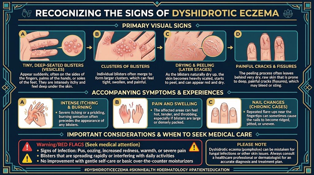
The initial warning sign often arrives as a sudden cluster of deep-seated, tapioca-like blisters, typically erupting along the sides of your fingers or the edges of your palms. These tiny vesicles, measuring 1-2 millimeters in diameter, sit deeper within the skin than surface-level irritation, giving the affected area a firm, bumpy texture rather than a delicate, fluid-filled blister that breaks easily. A relentless, prickling itch characterizes this first phase, frequently preceding any visible change by several hours, a phenomenon dermatologists describe as a prodromal sensation. Your skin between the blisters may appear entirely normal at first, though a closer look often reveals a faint, reddish undertone that intensifies as the episode progresses over the following 12 to 24 hours.
Persistent scratching or spontaneous rupture transforms the initial presentation into a more complex, scaly landscape. The vesicles eventually merge, burst, and weep a clear serous fluid, which can crust into yellowish, honey-colored layers that mimic secondary bacterial impetigo. Swelling across the palm or the arch of the foot creates a tight, uncomfortable stiffness that limits joint mobility, making simple tasks like buttoning a shirt unexpectedly difficult. After the weeping stage subsides, typically within two to three weeks, the affected skin dries out dramatically, revealing deep, linear cracks and fissures that radiate along the natural flexion lines of your hands. These painful splits can bleed with movement and serve as entry points for bacterial infections, a complication that turns a self-limiting flare into a clinical problem requiring antibiotic intervention.
Chronic, untreated cycles distort the skin’s architecture in distinct patterns invisible during an initial outbreak. Your skin exhibits lichenification, a thick, leathery reinforcement of the epidermis that develops in response to months of inflammatory signaling and unconscious rubbing. The normal fingerprint ridges disappear under a glossy, smooth surface, only to be replaced later by irregular scaling and peeling in circular patches. A hallmark of the vesicular drying phase involves the formation of large, brownish, coin-shaped areas of hyperkeratosis where the blisters once sat, a residue that lingers long after the itching subsides. Nail involvement presents concurrently in approximately 10% of persistent cases, manifesting as transverse ridging, thickening, or irregular pitting of the fingernail plates. Swollen lymph nodes in the affected limb signal a spreading infection, an escalation that demands immediate attention.
- Blisters contain clear fluid, not pus, unless a secondary infection has developed.
- Peeling often occurs 3 to 4 weeks after the initial blister eruption, even without scratching.
- Stress-induced flares may appear within 48 hours of a triggering emotional event.
- Metal sensitivity, particularly to nickel or cobalt, can reproduce identical blisters in a localized patch test.
- A potassium hydroxide skin scraping helps differentiate the blisters from a fungal infection.
Dyshidrotic eczema on the hands
Hand involvement concentrates heavily on the volar surfaces, targeting the palms and the lateral aspects of the digits with a precision that spares the dorsal skin. Blisters emerge in a characteristic distribution along the sides of your index and middle fingers first, coalescing into larger, multi-chambered bullae as the inflammatory cascade intensifies. The deep dermal location of these vesicles within the thick stratum corneum of your palm means they do not rupture easily; instead, they often persist as firm, painful nodules that interfere with your grip strength and tactile sensitivity. Occupational activities involving repetitive friction, such as prolonged writing, using hand tools, or gripping a steering wheel for hours, exacerbate the fluid accumulation and accelerate the weeping phase within days of the initial outbreak.
Fissuring across the palmar creases represents the most debilitating progression of hand lesions, a direct consequence of the skin’s attempt to shed the inflamed epidermal layers. These cracks, which can reach the dermal layer, bleed upon extension of the hand and create a cycle of pain and restricted movement that leads to muscle guarding in the forearm. Applying any pressure, such as opening a jar or lifting a weight, forces the edges of the fissures further apart, delaying closure by days. Your sleep quality deteriorates accordingly, as the nocturnal drop in cortisol amplifies both the itching and the throbbing from deep tissue inflammation. Secondary allergic contact dermatitis frequently complicates the clinical picture because the compromised skin barrier allows common sensitizers like fragrances and preservatives in soaps to penetrate deeply.
Recurrent episodes carve a distinct fingerprint of damage onto the nail apparatus and surrounding tissue, changing the hand’s appearance permanently in some individuals. Chronic paronychia develops as the proximal nail fold becomes swollen and detached from the nail plate, allowing moisture and yeast to colonize the subungual space. The nail plate responds with irregular pitting, a roughened surface texture, and a brownish-yellow discoloration that can be mistaken for a fungal infection. Longitudinal ridging and Beau’s lines, horizontal grooves that record the timeline of an acute flare, become visible months after the skin has healed. Approximately one in five patients with hand eczema report a subsequent loss of full range of motion in the proximal interphalangeal joints due to the cumulative effects of disuse and soft tissue contracture.
Dyshidrotic eczema on the feet
Foot involvement localizes to the medial arch and the plantar surface, regions rich in eccrine sweat glands where the occlusive environment inside footwear creates a uniquely moist microenvironment. The blisters on your instep tend to be larger and more isolated than their hand counterparts, often reaching 5 millimeters in diameter before they rupture, releasing a sticky, serum-rich fluid that soaks into socks. A burning, rather than purely itching, sensation dominates the sensory experience because the thick plantar skin traps inflammatory exudate under pressure with every weight-bearing step. The natural warmth and humidity inside athletic shoes or work boots accelerate the vesicle formation, causing a single friction point to spawn a cluster of 10 or more blisters within an afternoon of intense walking or standing.
Maceration sets in rapidly once the blisters rupture, turning the surrounding skin white, wrinkled, and highly vulnerable to bacterial ingress. Pseudomonas aeruginosa, recognizable by a greenish-blue discoloration on socks, colonizes the macerated tissue and produces a distinctive, sweetish odor. The interdigital spaces between your toes become sodden and peel in sheets, mimicking the presentation of tinea pedis so closely that misdiagnosis occurs in roughly half of the cases initially seen in primary care. Fungal elements, however, are absent on a KOH preparation, and the distribution remains bilateral and symmetrical, favoring the arches rather than the toe webs. Walking barefoot on hard surfaces becomes excruciating as the dermal papillae, stripped of their protective keratin layer, register every grain of sand with amplified sensitivity.
Persistent foot involvement often triggers a distinctive pattern of lymphangitic streaking up the ankle, a red line that tracks the drainage of inflammatory mediators along the superficial lymphatic channels. Chronic lymphedema of the dorsum thickens the skin over the metatarsal heads, creating a cobblestone appearance that specialists recognize as elephantiasis nostras verrucosa in advanced cases. Hyperhidrosis, present in up to 40% of individuals with pedal dyshidrotic eczema, supplies a continuous source of moisture that perpetuates the cycle of blistering and maceration. Ionizing the damp environment with aluminum chloride preparations or targeted botulinum injections reduces sweat output, but the thickened, fissured skin on the heel takes six to eight weeks to regenerate fully after the active eruption subsides.
Can it affect areas beyond the hands and feet?
The designation of dyshidrotic eczema as a palmoplantar condition reflects its overwhelming predilection for these sites, yet isolated reports confirm a phenomenon known as an “id reaction” that involves distant skin regions. An autoeczematous response can spread the inflammation to your forearms, upper chest, and even the lateral aspects of your neck when the primary eruption on the hands or feet is particularly severe and left untreated. These secondary patches do not contain the deep-seated tapioca vesicles of dyshidrosis; instead, they present as diffuse, erythematous, and intensely pruritic plaques that mimic a generalized eczematous dermatitis. Dermatologists identify this pattern through its temporal relationship to a virulent foot or hand flare, appearing 7 to 10 days after the primary blisters reach their peak intensity.
Nail changes on the toes can mirror the hand involvement, creating a widespread dystrophy that makes the 20-nail assessment a critical part of the clinical examination. All ten fingernails and ten toenails may exhibit onycholysis, with the nail plate lifting from the bed starting at the distal edge, a process that happens silently until debris accumulates beneath the loose nail. Yellowish-brown discoloration and subungual hyperkeratosis build a thick, crumbly material that often traps moisture and promotes the growth of secondary Candida species. The periorbital skin, though rarely involved directly, can swell in a hypersensitivity response during severe generalized flares, adding a cosmetic dimension that increases emotional distress and disrupts facial recognition software in daily digital interactions.
Rare acral extensions migrate the blistering pattern onto the dorsal surfaces of the wrists and the ankles, areas where the skin is thinner and more prone to oozing. These extensions challenge the conventional diagnostic boundaries and often prompt a broader workup that includes patch testing for systemic contact allergens like ingested nickel or cobalt salts. A low-nickel diet, reducing intake of cocoa, legumes, and canned foods, has resolved recalcitrant, widespread eruptions in a subset of patients whose standard topical treatments had failed for over a year. Pediatric cases, though accounting for less than 5% of all dyshidrotic eczema presentations, sometimes manifest as bullous impetigo mimics on the dorsal toes, complete with annular, crusted plaques that swab negative for staphylococcal or streptococcal antigens.
Treatment and Holistic Approaches
Conventional treatment options
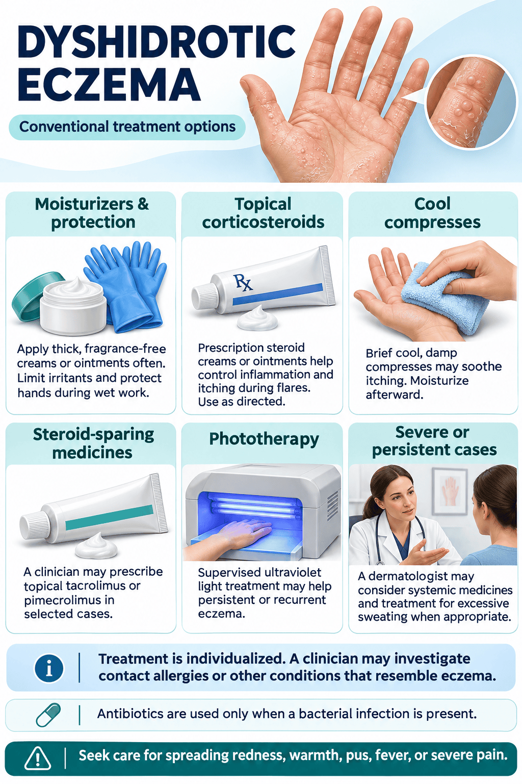
Your dermatologist can map out a treatment path that corresponds to the eruption phase of your vesicles. A first-line strategy during acute blistering involves high-potency topical corticosteroids, such as clobetasol propionate ointment, applied twice daily under occlusion to penetrate the thickened palmar or plantar skin. Soaks using aluminum acetate solution, prepared from Domeboro packets and cool water, and dries the weeping blisters through astringent action over a 15-minute immersion. When the attack is severe enough to disrupt sleep due to itching, a short oral prednisone taper starting at 40 mg and reducing over ten days provides systemic relief, though the rebound phenomenon often sparks a new crop of vesicles within a week of discontinuation. Wetter lesions may signal a secondary staphylococcal infection, warranting a bacterial culture and a course of cephalexin before steroid potency is increased.
| Treatment Tier | Example Agent | Key Application Detail |
|---|---|---|
| Topical corticosteroid | Clobetasol propionate 0.05% | Twice daily under plastic wrap occlusion |
| Calcineurin inhibitor | Tacrolimus 0.1% ointment | Rotate with steroids to limit skin atrophy. |
| Systemic immunomodulator | Methotrexate 15 mg weekly | Reserved for chronic refractory cases |
Calcineurin inhibitors like tacrolimus 0.1% ointment offer a steroid-sparing bridge for the chronic desquamative phase, rotated on alternate days to protect the epidermis from atrophy. Physicians sometimes prescribe alitretinoin at 30 mg daily, an oral retinoid available in Canada and several European nations under the name Toctino, which achieved clearance in roughly half of patients with severe chronic hand eczema within 12 to 24 weeks during its pivotal trial. For recalcitrant flares that resist these measures, narrow-band UVB phototherapy administered three times weekly in a dermatology office can slow keratinocyte proliferation, and botulinum toxin injections into the palms have temporarily interrupted sweating in a small series of patients with hyperhidrosis-driven pompholyx.
Dysport or Botox injections blocking eccrine sweat glands represent an emerging intervention when palmar hyperhidrosis is a clear trigger, though the procedure carries a dose-dependent risk of transient grip weakness. Potassium permanganate soaks, diluted to a pale pink 1:10,000 concentration, dry blisters in settings where aluminum acetate is unavailable. A dermatologist at the National Eczema Association’s 2023 clinical symposium emphasized combining an induction phase of medication with a maintenance protocol of barrier repair to reduce the cumulative steroid burden, a stance now reflected in step-down treatment algorithms across major eczema centers.
Natural and holistic approaches
Soaking 10 minutes nightly in a basin of cool water laced with a handful of whole oats, contained inside a muslin bag to prevent drain clogging, calms prickling heat through beta-glucan deposition on the skin surface. Sun protection becomes non-negotiable once the blisters dry and crack, because UV-exposed fissures heal with deeper post-inflammatory hyperpigmentation that can linger six months or longer. A zinc oxide paste applied to open cracks before bed creates a flexible seal, and a pair of thin cotton gloves worn overnight locks the barrier in place while you sleep. Compresses using chilled black tea, steeped until dark and pressed to the palms for 20 minutes twice daily, leverage the astringent tannic acid to tighten weeping vesicles much like an aluminum acetate preparation, a method documented in European dermatology texts for over a century.
Strategic avoidance of nickel and cobalt in the diet makes a measurable difference for a subgroup of dyshidrotic patients with systemic contact allergy confirmed by patch testing. High-nickel foods suspended during a clearance protocol include canned legumes, chocolate, whole-grain oatmeal, and soy products, while cobalt-rich items such as flax seeds and Brazil nuts are paused on a parallel track. Reintroduction after a 28-day elimination period reveals individual thresholds, with some patients reporting that a single square of dark chocolate triggers a new crop of vesicles within 48 hours. Cold-pressed sunflower seed oil massaged into peeling skin delivers linoleic acid directly to a ceramide-depleted barrier, and its low comedogenic rating prevents the pore occlusion that heavier plant butters can cause on the dorsal foot.
Hypochlorous acid spray, available medical-grade in concentrations of 0.02%, mists onto itching palms throughout the day to lower bacterial colonization without the stinging alcohol sensation of traditional sanitizers. A sleep protocol elevating the affected hands on a folded pillow while maintaining bedroom humidity at 40 percent with a cool-mist humidifier addresses the nocturnal pruritus surge rooted in circadian cortisol dips. Acupuncture targeting LI4 and LI11 points showed a statistically significant reduction in visual analog scale itch scores in a 2022 pilot study of 45 pompholyx patients at a Shanghai teaching hospital, an approach that some integrative dermatologists now incorporate as an adjunct during steroid tapering.
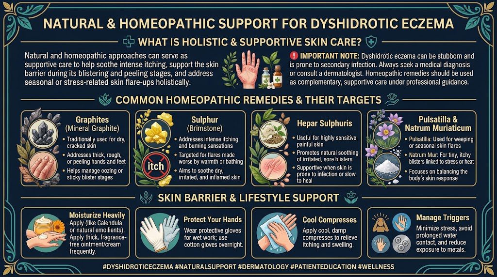
Homeopathic remedies
Practitioners of homeopathy generally select remedies based on an individual’s overall symptom pattern rather than the condition alone. Graphites 30C remains the most frequently indicated polychrest for the fissured, sticky discharge stage of pompholyx, prescribed when the skin cracks are deep enough to bleed on extension of the fingers and a honey-like exudate glues the skin folds upon waking. Rhus toxicodendron 30C aligns with a very different picture: relentless restlessness and tingling that compels you to rub the palms on rough fabrics for temporary relief, a presentation that worsens in cold damp weather and improves markedly after running the hands under water as hot as tolerated. A constitutional prescribing session with a board-certified homeopath can place the hand eruption within the context of your overall reactive pattern, linking mental stressors to the timing of flares in a way that a topical-only strategy cannot capture.
Dosing typically follows a 4-hour interval during the acute weeping stage, tapering to twice daily as the blisters transition to dry scaling, with the remedy pellets dissolved under the tongue at least 15 minutes away from food or mint toothpaste. Petroleum 6X trituration tablets, taken three times daily while fissures remain deep enough to catch on silk, address the severe dryness phase that persists long after the blisters have resolved. The homeopathic analysis of a 2021 case series published in Complementary Therapies in Medicine documented 11 of 14 dyshidrotic patients achieving at least 50 percent clearance on a physician global assessment after three months of individualized prescribing, though the mechanism proposed rests on a biophysical signaling model distinct from ligand-receptor pharmacology.
Natrum muriaticum 200C, given as a single dose and left to act over a month, suits the emotional reserve pattern where grief or disappointment preceded the onset of pearl-like blisters along the lateral fingers, often in a person who craves salt and avoids direct sun exposure. A combination approach that layers this constitutional dose atop the daily Petroleum 6X can bridge the gap between acute symptom suppression and deeper rebalancing, though any response lasting less than six weeks prompts a reassessment of the remedy match. Sulphur 30C applied sparingly, usually once weekly, covers the intensely burning, red vesicles that worsen with wool contact and washing, though its use demands caution because a premature repeat dose can provoke a temporary aggravation of the very itching you aim to quiet.
Final Words
Dyshidrotic eczema presents a distinct clinical picture with its deep-seated, tapioca-like vesicles erupting primarily along the lateral aspects of fingers, palms, and soles. A dermatologist in a typical urban clinic might confirm half a dozen new cases each week during peak allergy seasons, underscoring how common this condition really is. The intense pruritus often arrives before any visible skin change, a prodromal signal that distinguishes this form of dermatitis from other hand eczemas. Once the blisters resolve, the subsequent drying phase leaves fissures that crack painfully with every hand movement, creating a cycle of compromised barrier function and environmental insult.
The multifactorial origin means a single “cure” remains elusive, yet targeted management yields substantial relief. Nickel and cobalt ingestion, even from foods like oats and chocolate in sensitized individuals, can perpetuate flares just as effectively as direct contact with costume jewelry or wet work. Stress-induced spikes in cortisol weaken the skin’s antimicrobial defenses, and a patient tracking personal outbreaks against a life-event calendar will frequently map exacerbations to periods of sleep deprivation or deadline pressure. An integrative protocol that combines a high-potency topical corticosteroid during the acute vesicular phase with a heavy, ceramide-rich emollient during the cracked recovery period addresses both ends of the inflammatory arc.
Long-term control hinges on habit modification and trigger avoidance rather than perpetual pharmaceutical suppression. Individuals who wear cotton liners beneath vinyl or nitrile gloves for all wet chores—from dishwashing to infant bathing—report a measurable reduction in cyclic recurrence. A systematic elimination protocol starting with the most common allergenic metals, followed by a supervised reintroduction phase, provides a personalized map of dietary thresholds rather than a generic restriction list. The evidence from stress-reduction interventions, particularly heart rate variability biofeedback training, indicates that dampening the sympathetic overdrive directly correlates with fewer vesicles per episode. By mapping your own trigger profile and deploying a staged response—from barrier repair to immunomodulation—you reclaim agency over a condition that thrives on inattention.
FAQ

How much does dyshidrotic eczema treatment cost?
Costs range from under $20 for over-the-counter emollients and hydrocortisone to several hundred dollars for prescription topicals, phototherapy, or specialist visits, depending on insurance and treatment intensity. The biggest variable is whether stronger medications like calcineurin inhibitors or systemic drugs are required.
How long does it take for dyshidrotic eczema blisters to heal?
Blisters usually dry up within two to three weeks with consistent management, though the skin may remain tender and prone to cracking for several more weeks. Full barrier repair can take over a month if there are deep fissures or secondary infections.
What are the risks of not treating dyshidrosis?
Untreated dyshidrosis can lead to painful skin fissures, secondary bacterial or fungal infections, and chronic thickening of the skin that prolongs discomfort. The main risk is recurrent cellulitis requiring antibiotics when staph bacteria enter through open cracks.
What mistakes make dyshidrotic eczema worse?
Frequent hand washing with hot water and harsh soaps, wearing occlusive gloves for long periods, and scratching or popping blisters all aggravate the condition. The most common error is overusing topical steroids on thin skin without periodic breaks, which can lead to atrophy and rebound flares.
Are there alternatives to steroid creams for dyshidrotic eczema?
Yes, alternatives include calcineurin inhibitors (tacrolimus, pimecrolimus), barrier repair moisturizers with ceramides, dilute vinegar soaks to dry blisters, and phototherapy. Coal tar preparations may also help for chronic, thickened lesions, though they are less cosmetically acceptable.
Is holistic treatment worth trying for blistered hands and feet?
Holistic approaches like cold compresses, aloe vera, calendula creams, and stress reduction often provide significant relief of dyshidrotic eczema, especially for mild cases or as complementary support alongside prescribed treatments. They are generally worth a trial because they carry low risk, but severe flares still require medical evaluation.
Can homeopathic remedies help with dyshidrotic eczema?
Many people report symptomatic relief with homeopathic preparations like Graphites or Sulphur chosen to match their individual skin pattern, though no reproducible clinical evidence proves efficacy for dyshidrotic eczema beyond placebo. Their main value is as part of a broader holistic plan that also addresses triggers and skin care habits.
Natural Treatments for Dyshidrotic Eczema in Philadelphia
Dyshidrotic Eczema is a serious medical condition that significantly decreases the degree of quality of life. While allopathic medicine lacks an answer for treating eczema, some holistic and naturopathic approaches have proven effective and safe. The natural eczema treatments may include the use of Homeopathy, Acupuncture, Chinese herbs, and, of course, an eczema diet. All these forms of natural eczema treatments are available at the Philadelphia Holistic Treatment, where licensed practitioners work under the supervision of Victor Tsan. To schedule your initial holistic evaluation with Victor Tsan, contact our clinic at (267) 403-3085 or use our online scheduling application.